Department of Pathology and Experimental Therapeutics, Bellvitge University Hospital-IDIBELL, 08908, Hospitalet de Llobregat, Spain.
Institute of Biomedicine (IBUB) of the University of Barcelona (UB), 08028, Barcelona, Spain.
Sci Rep. 2019 May 2;9(1):6811. doi: 10.1038/s41598-019-43080-2.
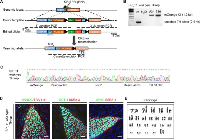
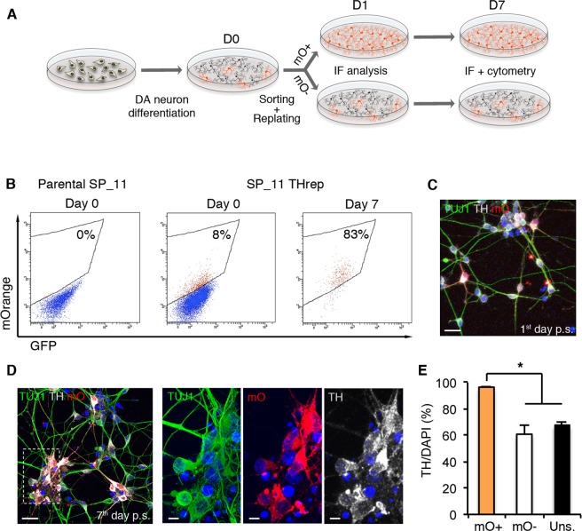
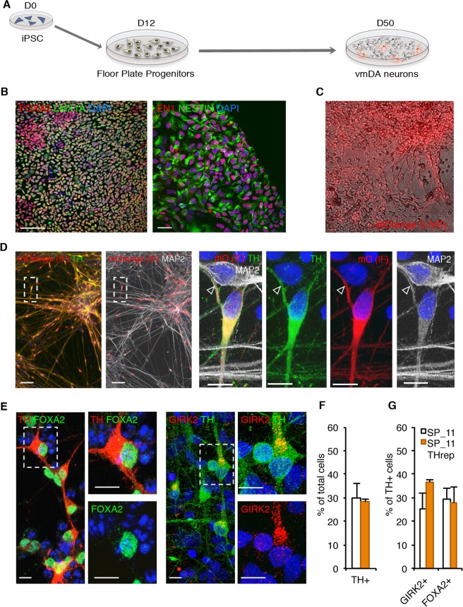
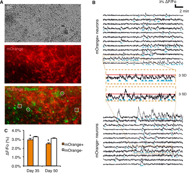
Patient-specific induced pluripotent stem cells (iPSCs) are a powerful tool to investigate the molecular mechanisms underlying Parkinson's disease (PD), and might provide novel platforms for systematic drug screening. Several strategies have been developed to generate iPSC-derived tyrosine hydroxylase (TH)-positive dopaminergic neurons (DAn), the clinically relevant cell type in PD; however, they often result in mixed neuronal cultures containing only a small proportion of TH-positive DAn. To overcome this limitation, we used CRISPR/Cas9-based editing to generate a human iPSC line expressing a fluorescent protein (mOrange) knocked-in at the last exon of the TH locus. After differentiation of the TH-mOrange reporter iPSC line, we confirmed that mOrange expression faithfully mimicked endogenous TH expression in iPSC-derived DAn. We also employed calcium imaging techniques to determine the intrinsic functional differences between dopaminergic and non-dopaminergic ventral midbrain neurons. Crucially, the brightness of mOrange allowed direct visualization of TH-expressing cells in heterogeneous cultures, and enabled us to isolate live mOrange-positive cells through fluorescence-activated cell sorting, for further differentiation. This technique, coupled to refined imaging and data processing tools, could advance the investigation of PD pathogenesis and might offer a platform to test potential new therapeutics for PD and other neurodegenerative diseases.
患者特异性诱导多能干细胞(iPSC)是研究帕金森病(PD)分子机制的有力工具,也可能为系统药物筛选提供新的平台。已经开发了几种策略来生成 iPSC 衍生的酪氨酸羟化酶(TH)阳性多巴胺能神经元(DAn),这是 PD 中临床相关的细胞类型;然而,它们通常会导致混合神经元培养物,其中仅包含一小部分 TH 阳性 DAn。为了克服这一限制,我们使用基于 CRISPR/Cas9 的编辑技术生成了一条表达荧光蛋白(mOrange)的人类 iPSC 系,该蛋白在 TH 基因座的最后外显子处被敲入。在 TH-mOrange 报告基因 iPSC 系分化后,我们证实 mOrange 表达在 iPSC 衍生的 DAn 中忠实地模拟了内源性 TH 表达。我们还采用钙成像技术来确定多巴胺能和非多巴胺能腹侧中脑神经元之间的内在功能差异。至关重要的是,mOrange 的亮度允许直接观察异质培养物中表达 TH 的细胞,并使我们能够通过荧光激活细胞分选分离活的 mOrange 阳性细胞,以进一步分化。这项技术与改良的成像和数据处理工具相结合,可以推进 PD 发病机制的研究,并可能为测试 PD 和其他神经退行性疾病的潜在新疗法提供一个平台。