Riihilä P, Viiklepp K, Nissinen L, Farshchian M, Kallajoki M, Kivisaari A, Meri S, Peltonen J, Peltonen S, Kähäri V-M
Department of Dermatology, University of Turku and Turku University Hospital, Hämeentie 11 TE6, FI-20520, Turku, Finland.
MediCity Research Laboratory, University of Turku, Turku, Finland.
Br J Dermatol. 2020 Mar;182(3):658-670. doi: 10.1111/bjd.18095. Epub 2019 Jul 28.
The incidence of epidermal keratinocyte-derived cutaneous squamous cell carcinoma (cSCC) is increasing worldwide.
To study the role of the complement classical pathway components C1q, C1r and C1s in the progression of cSCC.
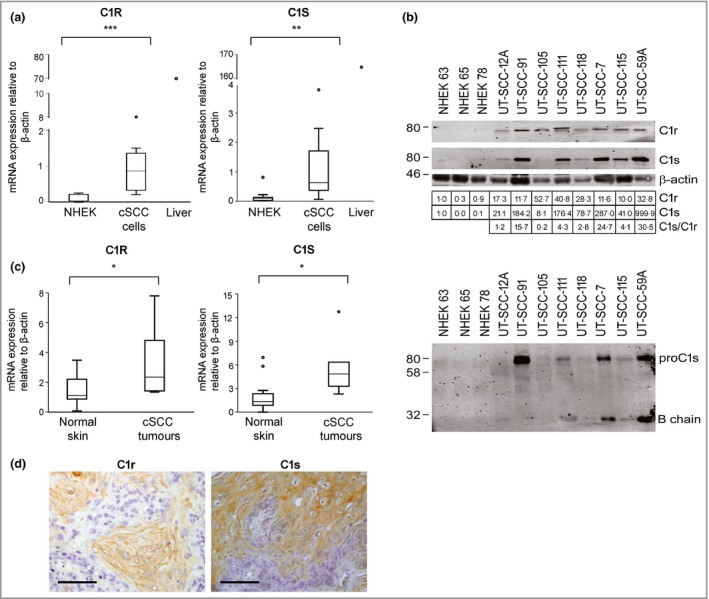
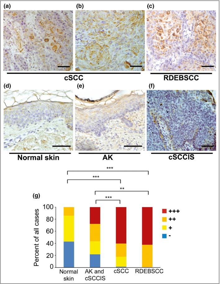
The mRNA levels of C1Q subunits and C1R and C1S in cSCC cell lines, normal human epidermal keratinocytes, cSCC tumours in vivo and normal skin were analysed with quantitative real-time polymerase chain reaction. The production of C1r and C1s was determined with Western blotting. The expression of C1r and C1s in tissue samples in vivo was analysed with immunohistochemistry and further investigated in human cSCC xenografts by knocking down C1r and C1s.
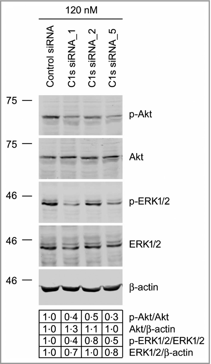
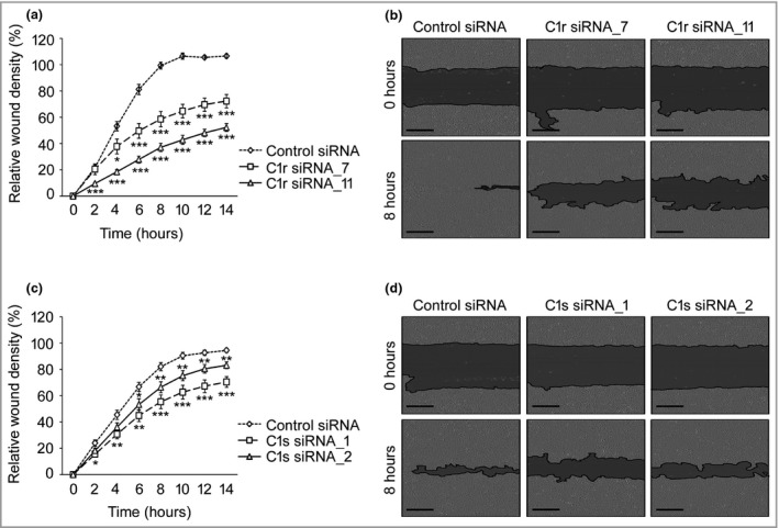
Significantly elevated C1R and C1S mRNA levels and production of C1r and C1s were detected in cSCC cells, compared with normal human epidermal keratinocytes. The mRNA levels of C1R and C1S were markedly elevated in cSCC tumours in vivo compared with normal skin. Abundant expression of C1r and C1s by tumour cells was detected in invasive sporadic cSCCs and recessive dystrophic epidermolysis bullosa-associated cSCCs, whereas the expression of C1r and C1s was lower in cSCC in situ, actinic keratosis and normal skin. Knockdown of C1r and C1s expression in cSCC cells inhibited activation of extracellular signal-related kinase 1/2 and Akt, promoted apoptosis of cSCC cells and significantly suppressed growth and vascularization of human cSCC xenograft tumours in vivo.
These results provide evidence for the role of tumour-cell-derived C1r and C1s in the progression of cSCC and identify them as biomarkers and putative therapeutic targets in cSCC. What's already known about this topic? The incidences of actinic keratosis, cutaneous squamous cell carcinoma (cSCC) in situ and invasive cSCC are increasing globally. Few specific biomarkers for progression of cSCC have been identified, and no biological markers are in clinical use to predict the aggressiveness of actinic keratosis, cSCC in situ and invasive cSCC. What does this study add? Our results provide novel evidence for the role of complement classical pathway components C1r and C1s in the progression of cSCC. What is the translational message? Our results identify complement classical pathway components C1r and C1s as biomarkers and putative therapeutic targets in cSCC.
表皮角质形成细胞来源的皮肤鳞状细胞癌(cSCC)的发病率在全球范围内呈上升趋势。
研究补体经典途径成分C1q、C1r和C1s在cSCC进展中的作用。
采用定量实时聚合酶链反应分析cSCC细胞系、正常人表皮角质形成细胞、体内cSCC肿瘤及正常皮肤中C1Q亚基、C1R和C1S的mRNA水平。用蛋白质免疫印迹法测定C1r和C1s的产生。用免疫组织化学法分析体内组织样本中C1r和C1s的表达,并通过敲低C1r和C1s在人cSCC异种移植瘤中进一步研究。
与正常人表皮角质形成细胞相比,在cSCC细胞中检测到C1R和C1S mRNA水平显著升高,且C1r和C1s产生增加。与正常皮肤相比,体内cSCC肿瘤中C1R和C1S的mRNA水平明显升高。在侵袭性散发性cSCC和隐性营养不良性大疱性表皮松解症相关的cSCC中检测到肿瘤细胞大量表达C1r和C1s,而原位cSCC、光化性角化病和正常皮肤中C1r和C1s的表达较低。敲低cSCC细胞中C1r和C1s的表达可抑制细胞外信号调节激酶1/2和Akt的激活,促进cSCC细胞凋亡,并显著抑制体内人cSCC异种移植瘤的生长和血管生成。
这些结果为肿瘤细胞来源的C1r和C1s在cSCC进展中的作用提供了证据,并将它们鉴定为cSCC的生物标志物和潜在治疗靶点。关于该主题已知的信息有哪些?光化性角化病、原位皮肤鳞状细胞癌(cSCC)和侵袭性cSCC的发病率在全球范围内都在增加。已鉴定出的用于cSCC进展的特异性生物标志物很少,且尚无生物标志物用于临床预测光化性角化病、原位cSCC和侵袭性cSCC的侵袭性。本研究增加了什么?我们的结果为补体经典途径成分C1r和C1s在cSCC进展中的作用提供了新的证据。转化信息是什么?我们的结果将补体经典途径成分C1r和C1s鉴定为cSCC的生物标志物和潜在治疗靶点。