• 文献检索
  • 文档翻译
  • 深度研究
  • 学术资讯
  • Suppr Zotero 插件Zotero 插件
  • 邀请有礼
  • 套餐&价格
  • 历史记录
应用&插件
Suppr Zotero 插件Zotero 插件浏览器插件Mac 客户端Windows 客户端微信小程序
定价
高级版会员购买积分包购买API积分包
服务
文献检索文档翻译深度研究API 文档MCP 服务
关于我们
关于 Suppr公司介绍联系我们用户协议隐私条款
关注我们

Suppr 超能文献

核心技术专利:CN118964589B侵权必究
粤ICP备2023148730 号-1Suppr @ 2026

文献检索

告别复杂PubMed语法,用中文像聊天一样搜索,搜遍4000万医学文献。AI智能推荐,让科研检索更轻松。

立即免费搜索

文件翻译

保留排版,准确专业,支持PDF/Word/PPT等文件格式,支持 12+语言互译。

免费翻译文档

深度研究

AI帮你快速写综述,25分钟生成高质量综述,智能提取关键信息,辅助科研写作。

立即免费体验

血管周围间隙液导致脑白质弥散张量成像改变。

Perivascular space fluid contributes to diffusion tensor imaging changes in white matter.

机构信息

Laboratory of Neuro Imaging, USC Mark and Mary Stevens Neuroimaging and Informatics Institute, Keck School of Medicine of USC, University of Southern California, Los Angeles, USA.

Laboratory of Neuro Imaging, USC Mark and Mary Stevens Neuroimaging and Informatics Institute, Keck School of Medicine of USC, University of Southern California, Los Angeles, USA.

出版信息

Neuroimage. 2019 Aug 15;197:243-254. doi: 10.1016/j.neuroimage.2019.04.070. Epub 2019 Apr 30.

DOI:10.1016/j.neuroimage.2019.04.070
PMID:31051291
原文链接:https://pmc.ncbi.nlm.nih.gov/articles/PMC6591070/
Abstract

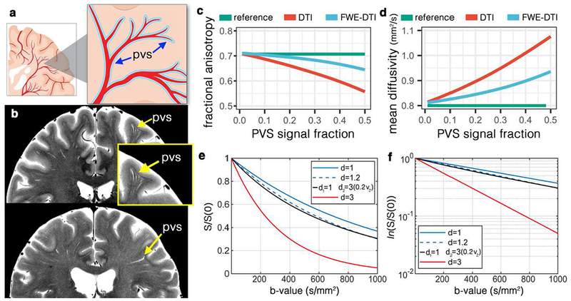
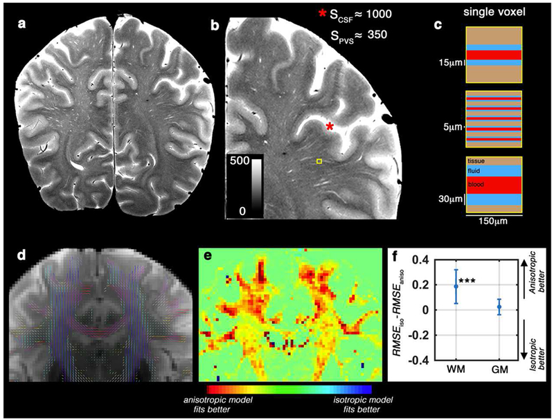
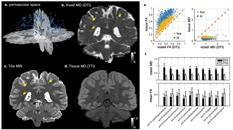
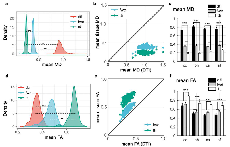
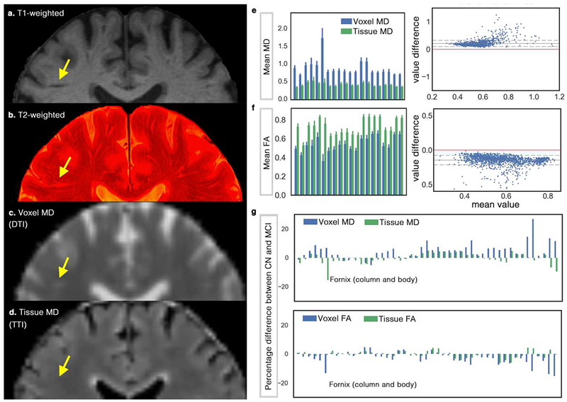
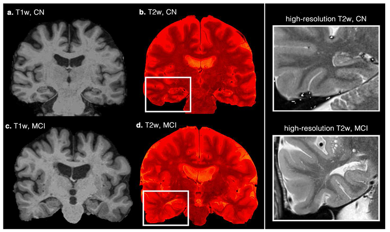
Diffusion tensor imaging (DTI) has been extensively used to map changes in brain tissue related to neurological disorders. Among the most widespread DTI findings are increased mean diffusivity and decreased fractional anisotropy of white matter tissue in neurodegenerative diseases. Here we utilize multi-shell diffusion imaging to separate diffusion signal of the brain parenchyma from non-parenchymal fluid within the white matter. We show that unincorporated anisotropic water in perivascular space (PVS) significantly, and systematically, biases DTI measures, casting new light on the biological validity of many previously reported findings. Despite the challenge this poses for interpreting these past findings, our results suggest that multi-shell diffusion MRI provides a new opportunity for incorporating the PVS contribution, ultimately strengthening the clinical and scientific value of diffusion MRI.

摘要

弥散张量成像(DTI)已广泛用于绘制与神经退行性疾病相关的脑组织变化图。在最广泛的 DTI 发现中,神经退行性疾病中的白质组织的平均弥散度增加和各向异性分数降低。在这里,我们利用多壳层扩散成像将脑实质的扩散信号与白质中的非实质流体分开。我们表明,血管周围空间(PVS)中的未结合各向异性水显著且系统地影响 DTI 测量值,这为许多先前报道的发现的生物学有效性提供了新的认识。尽管这对解释这些过去的发现提出了挑战,但我们的结果表明,多壳层扩散 MRI 为纳入 PVS 贡献提供了新的机会,最终增强了扩散 MRI 的临床和科学价值。

https://cdn.ncbi.nlm.nih.gov/pmc/blobs/1461/6591070/c69c64acbfb1/nihms-1528734-f0007.jpg
https://cdn.ncbi.nlm.nih.gov/pmc/blobs/1461/6591070/329dc5feab04/nihms-1528734-f0001.jpg
https://cdn.ncbi.nlm.nih.gov/pmc/blobs/1461/6591070/09d3cff41ddb/nihms-1528734-f0002.jpg
https://cdn.ncbi.nlm.nih.gov/pmc/blobs/1461/6591070/24d722051429/nihms-1528734-f0003.jpg
https://cdn.ncbi.nlm.nih.gov/pmc/blobs/1461/6591070/24f9e0b0bae9/nihms-1528734-f0004.jpg
https://cdn.ncbi.nlm.nih.gov/pmc/blobs/1461/6591070/8c600222f245/nihms-1528734-f0005.jpg
https://cdn.ncbi.nlm.nih.gov/pmc/blobs/1461/6591070/c2d5eadf1801/nihms-1528734-f0006.jpg
https://cdn.ncbi.nlm.nih.gov/pmc/blobs/1461/6591070/c69c64acbfb1/nihms-1528734-f0007.jpg

相似文献

1
Perivascular space fluid contributes to diffusion tensor imaging changes in white matter.血管周围间隙液导致脑白质弥散张量成像改变。
Neuroimage. 2019 Aug 15;197:243-254. doi: 10.1016/j.neuroimage.2019.04.070. Epub 2019 Apr 30.
2
Differences in Gaussian diffusion tensor imaging and non-Gaussian diffusion kurtosis imaging model-based estimates of diffusion tensor invariants in the human brain.基于高斯扩散张量成像和非高斯扩散峰度成像模型的人脑扩散张量不变量估计差异。
Med Phys. 2016 May;43(5):2464. doi: 10.1118/1.4946819.
3
Diffusion Tensor Imaging (DTI).扩散张量成像(DTI)
Methods Mol Biol. 2018;1718:103-116. doi: 10.1007/978-1-4939-7531-0_7.
4
MR Diffusion Tractography to Identify and Characterize Microstructural White Matter Tract Changes in Systemic Lupus Erythematosus Patients.磁共振扩散张量成像用于识别和表征系统性红斑狼疮患者脑白质微结构的改变
Acad Radiol. 2016 Nov;23(11):1431-1440. doi: 10.1016/j.acra.2016.03.019. Epub 2016 Oct 13.
5
Short-term white matter alterations in Alzheimer's disease characterized by diffusion tensor imaging.以扩散张量成像为特征的阿尔茨海默病短期白质改变
J Magn Reson Imaging. 2016 Mar;43(3):627-34. doi: 10.1002/jmri.25017. Epub 2015 Jul 30.
6
A multimodal MRI approach to identify and characterize microstructural brain changes in neuropsychiatric systemic lupus erythematosus.一种用于识别和表征神经精神性系统性红斑狼疮患者脑微结构变化的多模态磁共振成像方法。
Neuroimage Clin. 2015 May 16;8:337-44. doi: 10.1016/j.nicl.2015.05.002. eCollection 2015.
7
Comparing a diffusion tensor and non-tensor approach to white matter fiber tractography in chronic stroke.比较扩散张量成像和非张量成像方法在慢性卒中白质纤维束成像中的应用
Neuroimage Clin. 2015 Mar 14;7:771-81. doi: 10.1016/j.nicl.2015.03.007. eCollection 2015.
8
Hemispheric Regional Based Analysis of Diffusion Tensor Imaging and Diffusion Tensor Tractography in Patients with Temporal Lobe Epilepsy and Correlation with Patient outcomes.基于半球区域的分析弥散张量成像和弥散张量纤维束成像在颞叶癫痫患者中的应用及其与患者预后的相关性。
Sci Rep. 2019 Jan 18;9(1):215. doi: 10.1038/s41598-018-36818-x.
9
White Matter Abnormalities in Multiple Sclerosis Evaluated by Quantitative Synthetic MRI, Diffusion Tensor Imaging, and Neurite Orientation Dispersion and Density Imaging.基于定量合成 MRI、弥散张量成像和神经丝取向弥散和密度成像评估多发性硬化的脑白质异常。
AJNR Am J Neuroradiol. 2019 Oct;40(10):1642-1648. doi: 10.3174/ajnr.A6209. Epub 2019 Sep 12.
10
Neuroimaging findings related to glymphatic system alterations in older adults with metabolic syndrome.与患有代谢综合征的老年人淋巴系统改变相关的神经影像学研究结果。
Neurobiol Dis. 2023 Feb;177:105990. doi: 10.1016/j.nbd.2023.105990. Epub 2023 Jan 5.

引用本文的文献

1
An exploratory study of the diffusion tensor imaging analysis along perivascular spaces (DTI-ALPS) index combined with quantitative analysis of choroid plexus volume and perivascular spaces in different cognitive stages of cerebral small vessel disease.一项关于沿血管周围间隙扩散张量成像分析(DTI-ALPS)指数联合脉络丛体积及血管周围间隙定量分析在脑小血管病不同认知阶段的探索性研究。
Quant Imaging Med Surg. 2025 Sep 1;15(9):8173-8188. doi: 10.21037/qims-2025-733. Epub 2025 Aug 15.
2
Perivascular and parenchymal fluid characteristics are related to age and cognitive performance across the lifespan.血管周围和实质液体特征与整个生命周期中的年龄和认知表现相关。
Imaging Neurosci (Camb). 2025 Apr 29;3. doi: 10.1162/imag_a_00549. eCollection 2025.
3

本文引用的文献

1
Flow of cerebrospinal fluid is driven by arterial pulsations and is reduced in hypertension.脑脊液的流动是由动脉搏动驱动的,在高血压时会减少。
Nat Commun. 2018 Nov 19;9(1):4878. doi: 10.1038/s41467-018-07318-3.
2
The glymphatic pathway in neurological disorders.神经疾病中的糖酵解途径。
Lancet Neurol. 2018 Nov;17(11):1016-1024. doi: 10.1016/S1474-4422(18)30318-1.
3
Rapid solution of the Bloch-Torrey equation in anisotropic tissue: Application to dynamic susceptibility contrast MRI of cerebral white matter.各向异性组织中 Bloch-Torrey 方程的快速求解:在脑白质动态磁化率对比 MRI 中的应用。
Testing retrogenesis and physiological explanations for tract-wise white matter aging: links to developmental order, fiber calibre, and vascularization.测试逆向发生及白质束老化的生理学解释:与发育顺序、纤维直径及血管形成的联系
Geroscience. 2025 Jul 15. doi: 10.1007/s11357-025-01773-9.
4
White Matter Geometry Confounds Diffusion Tensor Imaging Along Perivascular Space (DTI-ALPS) Measures.白质几何结构混淆了沿血管周围间隙的扩散张量成像(DTI-ALPS)测量结果。
Hum Brain Mapp. 2025 Jul;46(10):e70282. doi: 10.1002/hbm.70282.
5
The relationship of white matter tract orientation to vascular geometry in the human brain.人脑白质纤维束方向与血管几何结构的关系。
Sci Rep. 2025 May 26;15(1):18396. doi: 10.1038/s41598-025-99724-z.
6
Altered Clearance in Alzheimer's Disease and Cerebral Amyloid Angiopathy.阿尔茨海默病和脑淀粉样血管病中的清除率改变
Neuroimaging Clin N Am. 2025 May;35(2):277-286. doi: 10.1016/j.nic.2024.12.004. Epub 2025 Feb 19.
7
Diffusion Tensor Imaging in Neurofluids.神经流体中的扩散张量成像
Neuroimaging Clin N Am. 2025 May;35(2):211-222. doi: 10.1016/j.nic.2024.11.001. Epub 2025 Feb 21.
8
The relationship of white matter tract orientation to vascular geometry in the human brain.人类大脑中白质束方向与血管几何形状的关系。
bioRxiv. 2025 Mar 6:2025.03.06.641646. doi: 10.1101/2025.03.06.641646.
9
Modulation and distribution of extracellular free water and tract deficits in rhesus macaques before and after the initiation of emtricitabine + tenofovir disoproxil fumarate + dotutegravir treatment.恩曲他滨+替诺福韦酯富马酸盐+多替拉韦治疗开始前后恒河猴细胞外游离水的调节与分布及神经束缺损情况
Front Immunol. 2025 Feb 28;16:1463434. doi: 10.3389/fimmu.2025.1463434. eCollection 2025.
10
Association of concussion history with psychiatric symptoms, limbic system structure, and kynurenine pathway metabolites in healthy, collegiate-aged athletes.健康的大学适龄运动员中脑震荡病史与精神症状、边缘系统结构及犬尿氨酸途径代谢物的关联
Brain Behav Immun. 2025 Jan;123:619-630. doi: 10.1016/j.bbi.2024.10.015. Epub 2024 Oct 14.
Neuroimage. 2019 Jan 15;185:198-207. doi: 10.1016/j.neuroimage.2018.10.035. Epub 2018 Oct 15.
4
Quantifying brain microstructure with diffusion MRI: Theory and parameter estimation.用扩散 MRI 量化脑微观结构:理论与参数估计。
NMR Biomed. 2019 Apr;32(4):e3998. doi: 10.1002/nbm.3998. Epub 2018 Oct 15.
5
Non-invasive imaging of CSF-mediated brain clearance pathways via assessment of perivascular fluid movement with diffusion tensor MRI.应用扩散张量 MRI 评估血管周围液流动对脑脊液介导的脑内清除途径的无创性成像。
Elife. 2018 Jul 31;7:e34028. doi: 10.7554/eLife.34028.
6
Understanding the role of the perivascular space in cerebral small vessel disease.了解血管周围间隙在脑小血管疾病中的作用。
Cardiovasc Res. 2018 Sep 1;114(11):1462-1473. doi: 10.1093/cvr/cvy113.
7
Tau and neuroinflammation: What impact for Alzheimer's Disease and Tauopathies?tau 与神经炎症:对阿尔茨海默病和 tau 病有何影响?
Biomed J. 2018 Feb;41(1):21-33. doi: 10.1016/j.bj.2018.01.003. Epub 2018 Mar 20.
8
Impact of time-of-day on diffusivity measures of brain tissue derived from diffusion tensor imaging.基于扩散张量成像的脑组织结构各向异性弥散系数的时间依赖性研究
Neuroimage. 2018 Jun;173:25-34. doi: 10.1016/j.neuroimage.2018.02.026. Epub 2018 Feb 16.
9
Perivascular Spaces Segmentation in Brain MRI Using Optimal 3D Filtering.基于最优 3D 滤波的脑 MRI 血管周围空间分割。
Sci Rep. 2018 Feb 1;8(1):2132. doi: 10.1038/s41598-018-19781-5.
10
The pathology and pathophysiology of vascular dementia.血管性痴呆的病理学和病理生理学。
Neuropharmacology. 2018 May 15;134(Pt B):226-239. doi: 10.1016/j.neuropharm.2017.12.030. Epub 2017 Dec 19.