• 文献检索
  • 文档翻译
  • 深度研究
  • 学术资讯
  • Suppr Zotero 插件Zotero 插件
  • 邀请有礼
  • 套餐&价格
  • 历史记录
应用&插件
Suppr Zotero 插件Zotero 插件浏览器插件Mac 客户端Windows 客户端微信小程序
定价
高级版会员购买积分包购买API积分包
服务
文献检索文档翻译深度研究API 文档MCP 服务
关于我们
关于 Suppr公司介绍联系我们用户协议隐私条款
关注我们

Suppr 超能文献

核心技术专利:CN118964589B侵权必究
粤ICP备2023148730 号-1Suppr @ 2026

文献检索

告别复杂PubMed语法,用中文像聊天一样搜索,搜遍4000万医学文献。AI智能推荐,让科研检索更轻松。

立即免费搜索

文件翻译

保留排版,准确专业,支持PDF/Word/PPT等文件格式,支持 12+语言互译。

免费翻译文档

深度研究

AI帮你快速写综述,25分钟生成高质量综述,智能提取关键信息,辅助科研写作。

立即免费体验

脑脊液的流动是由动脉搏动驱动的,在高血压时会减少。

Flow of cerebrospinal fluid is driven by arterial pulsations and is reduced in hypertension.

机构信息

Center for Translational Neuromedicine, University of Rochester Medical Center, Rochester, NY, 14642, USA.

Department of Neuroscience, University of Rochester Medical Center, Rochester, NY, 14642, USA.

出版信息

Nat Commun. 2018 Nov 19;9(1):4878. doi: 10.1038/s41467-018-07318-3.

DOI:10.1038/s41467-018-07318-3
PMID:30451853
原文链接:https://pmc.ncbi.nlm.nih.gov/articles/PMC6242982/
Abstract

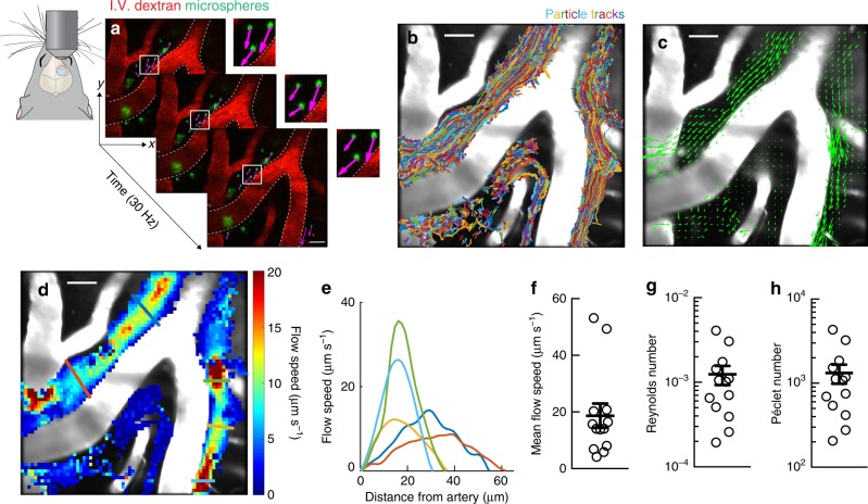
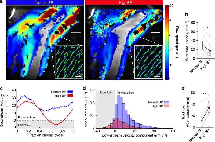
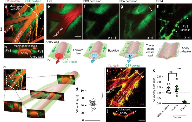
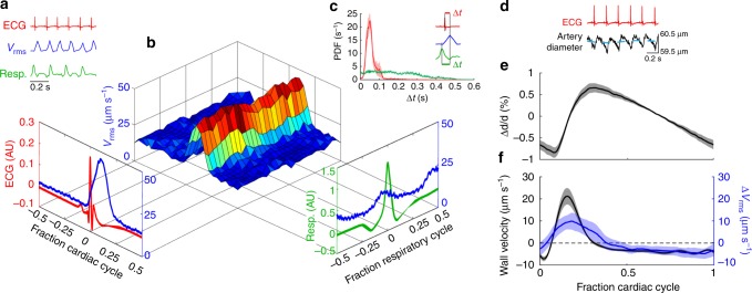
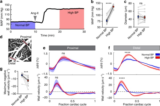
Flow of cerebrospinal fluid (CSF) through perivascular spaces (PVSs) in the brain is important for clearance of metabolic waste. Arterial pulsations are thought to drive flow, but this has never been quantitatively shown. We used particle tracking to quantify CSF flow velocities in PVSs of live mice. CSF flow is pulsatile and driven primarily by the cardiac cycle. The speed of the arterial wall matches that of the CSF, suggesting arterial wall motion is the principal driving mechanism, via a process known as perivascular pumping. Increasing blood pressure leaves the artery diameter unchanged but changes the pulsations of the arterial wall, increasing backflow and thereby reducing net flow in the PVS. Perfusion-fixation alters the normal flow direction and causes a 10-fold reduction in PVS size. We conclude that particle tracking velocimetry enables the study of CSF flow in unprecedented detail and that studying the PVS in vivo avoids fixation artifacts.

摘要

脑脊液(CSF)在脑内的血管周围间隙(PVS)中的流动对于清除代谢废物很重要。人们认为动脉搏动驱动了这种流动,但这从未被定量证明过。我们使用粒子追踪技术来量化活体小鼠 PVS 中的 CSF 流速。CSF 流动是脉动的,主要由心动周期驱动。动脉壁的速度与 CSF 的速度相匹配,这表明动脉壁的运动是主要的驱动机制,通过一种称为血管周围泵送的过程。增加血压不会改变动脉直径,但会改变动脉壁的搏动,增加逆流,从而减少 PVS 中的净流量。灌注固定会改变正常的流动方向,并使 PVS 大小减少 10 倍。我们的结论是,粒子追踪速度测量法使 CSF 流动的研究能够达到前所未有的细节水平,并且体内研究 PVS 可以避免固定伪影。

https://cdn.ncbi.nlm.nih.gov/pmc/blobs/3971/6242982/497b8e9787eb/41467_2018_7318_Fig5_HTML.jpg
https://cdn.ncbi.nlm.nih.gov/pmc/blobs/3971/6242982/3b5b0af3a218/41467_2018_7318_Fig1_HTML.jpg
https://cdn.ncbi.nlm.nih.gov/pmc/blobs/3971/6242982/d1b72b2ab5e3/41467_2018_7318_Fig2_HTML.jpg
https://cdn.ncbi.nlm.nih.gov/pmc/blobs/3971/6242982/92147028a203/41467_2018_7318_Fig3_HTML.jpg
https://cdn.ncbi.nlm.nih.gov/pmc/blobs/3971/6242982/73babc783e57/41467_2018_7318_Fig4_HTML.jpg
https://cdn.ncbi.nlm.nih.gov/pmc/blobs/3971/6242982/497b8e9787eb/41467_2018_7318_Fig5_HTML.jpg

相似文献

1
Flow of cerebrospinal fluid is driven by arterial pulsations and is reduced in hypertension.脑脊液的流动是由动脉搏动驱动的,在高血压时会减少。
Nat Commun. 2018 Nov 19;9(1):4878. doi: 10.1038/s41467-018-07318-3.
2
Arterial pulsation-driven cerebrospinal fluid flow in the perivascular space: a computational model.血管周围间隙中动脉搏动驱动的脑脊液流动:一种计算模型
Comput Methods Biomech Biomed Engin. 2003 Aug;6(4):235-41. doi: 10.1080/10255840310001606116.
3
The mechanisms behind perivascular fluid flow.血管周围液流的机制。
PLoS One. 2020 Dec 29;15(12):e0244442. doi: 10.1371/journal.pone.0244442. eCollection 2020.
4
Arterial pulsations drive oscillatory flow of CSF but not directional pumping.动脉搏动驱动 CSF 的振荡流动,但不驱动定向泵送。
Sci Rep. 2020 Jun 22;10(1):10102. doi: 10.1038/s41598-020-66887-w.
5
Arterial pulsation dependence of perivascular cerebrospinal fluid flow measured by dynamic diffusion tensor imaging in the human brain.通过动态扩散张量成像测量人脑血管周围脑脊液流动的动脉搏动依赖性。
Neuroimage. 2024 Aug 15;297:120653. doi: 10.1016/j.neuroimage.2024.120653. Epub 2024 May 23.
6
Arterial pulsation-dependent perivascular cerebrospinal fluid flow into the central canal in the sheep spinal cord.绵羊脊髓中动脉搏动依赖的血管周围脑脊液流入中央管。
J Neurosurg. 1997 Apr;86(4):686-93. doi: 10.3171/jns.1997.86.4.0686.
7
Phase offset between arterial pulsations and subarachnoid space pressure fluctuations are unlikely to drive periarterial cerebrospinal fluid flow.动脉搏动与蛛网膜下腔压力波动之间的相位偏移不太可能驱动动脉周围脑脊液流动。
Biomech Model Mechanobiol. 2021 Oct;20(5):1751-1766. doi: 10.1007/s10237-021-01474-0. Epub 2021 Jul 17.
8
In Vivo Imaging of Cerebrospinal Fluid Transport through the Intact Mouse Skull using Fluorescence Macroscopy.使用荧光显微镜对脑脊液通过完整小鼠颅骨的转运进行体内成像。
J Vis Exp. 2019 Jul 29(149). doi: 10.3791/59774.
9
Bulk flow of cerebrospinal fluid observed in periarterial spaces is not an artifact of injection.观察到的动脉周围腔隙中脑脊髓液的大体流动不是注射的假象。
Elife. 2021 Mar 9;10:e65958. doi: 10.7554/eLife.65958.
10
Cerebrospinal fluid flow driven by arterial pulsations in axisymmetric perivascular spaces: Analogy with Taylor's swimming sheet.动脉搏动驱动轴对称血管周围腔隙中脑脊液的流动:与泰勒游泳片的类比。
J Theor Biol. 2021 Aug 21;523:110709. doi: 10.1016/j.jtbi.2021.110709. Epub 2021 Apr 20.

引用本文的文献

1
Abnormal sleep blood pressure patterns are associated with the diffusion tensor imaging along the perivascular space index in cognitively impaired individuals.异常的睡眠血压模式与认知障碍个体沿血管周围间隙指数的扩散张量成像相关。
Front Aging Neurosci. 2025 Aug 29;17:1578270. doi: 10.3389/fnagi.2025.1578270. eCollection 2025.
2
Subacute Progression of Gait Disturbance and Consciousness Impairment Due to Communicating Hydrocephalus Associated With Vestibular Schwannoma.与前庭神经鞘瘤相关的交通性脑积水导致步态障碍和意识障碍的亚急性进展
Cureus. 2025 Aug 12;17(8):e89880. doi: 10.7759/cureus.89880. eCollection 2025 Aug.
3

本文引用的文献

1
Non-invasive imaging of CSF-mediated brain clearance pathways via assessment of perivascular fluid movement with diffusion tensor MRI.应用扩散张量 MRI 评估血管周围液流动对脑脊液介导的脑内清除途径的无创性成像。
Elife. 2018 Jul 31;7:e34028. doi: 10.7554/eLife.34028.
2
Magnetic resonance imaging provides evidence of glymphatic drainage from human brain to cervical lymph nodes.磁共振成像提供了从人脑到颈部淋巴结的糖质质淋巴引流的证据。
Sci Rep. 2018 May 8;8(1):7194. doi: 10.1038/s41598-018-25666-4.
3
2017 ACC/AHA/AAPA/ABC/ACPM/AGS/APhA/ASH/ASPC/NMA/PCNA Guideline for the Prevention, Detection, Evaluation, and Management of High Blood Pressure in Adults: A Report of the American College of Cardiology/American Heart Association Task Force on Clinical Practice Guidelines.
Uncloggable ventriculoperitoneal shunt system for hydrocephalus via an integrated soft robotic device: CLEARS device.
用于脑积水的通过集成软机器人装置实现的不可堵塞的脑室腹腔分流系统:CLEARS装置
Biomed Microdevices. 2025 Sep 11;27(3):41. doi: 10.1007/s10544-025-00769-8.
4
Arterial stiffness and vascular aging: mechanisms, prevention, and therapy.动脉僵硬度与血管衰老:机制、预防及治疗
Signal Transduct Target Ther. 2025 Sep 1;10(1):282. doi: 10.1038/s41392-025-02346-0.
5
Salvianolic Acid B and Ginsenoside Rg1 Combination Attenuates Cerebral Edema Accompanying Glymphatic Modulation.丹酚酸B与人参皂苷Rg1联合应用减轻伴随类淋巴系统调节的脑水肿。
Neurosci Bull. 2025 Aug 31. doi: 10.1007/s12264-025-01490-5.
6
Exploring cerebral small vessel disease, choroid plexus, demographics and vascular risk factors that affect glymphatic function.探索影响类淋巴系统功能的脑小血管疾病、脉络丛、人口统计学和血管危险因素。
Jpn J Radiol. 2025 Aug 27. doi: 10.1007/s11604-025-01843-0.
7
Alterations in MRI-visible perivascular spaces precede dementia diagnosis by 18 years in autosomal dominant Alzheimer's disease.在常染色体显性遗传性阿尔茨海默病中,磁共振成像可见的血管周围间隙改变比痴呆诊断提前18年出现。
Alzheimers Dement. 2025 Aug;21(8):e70588. doi: 10.1002/alz.70588.
8
Temporal fluctuation in lateral ventricle volume and its coupling with CSF inflow and global BOLD signal.侧脑室体积的时间波动及其与脑脊液流入和全脑血氧水平依赖信号的耦合。
Sci Rep. 2025 Aug 20;15(1):30537. doi: 10.1038/s41598-025-15842-8.
9
The Cerebral Lymphatic System: Function, Controversies, and Therapeutic Approaches for Central Nervous System Diseases.脑淋巴系统:中枢神经系统疾病的功能、争议及治疗方法
Cell Mol Neurobiol. 2025 Aug 19;45(1):80. doi: 10.1007/s10571-025-01598-2.
10
Morphology of the glymphatic and meningeal lymphatic structures of the bottlenose dolphin.宽吻海豚脑周淋巴系统和脑膜淋巴结构的形态学
Sci Rep. 2025 Aug 18;15(1):30216. doi: 10.1038/s41598-025-14840-0.
2017美国心脏病学会/美国心脏协会/美国医师协会/美国心脏病学学会/美国预防医学学院/美国老年病学会/美国药学协会/美国血液学会/美国预防医学学会/美国医学协会/美国初级保健医师学会成人高血压预防、检测、评估和管理指南:美国心脏病学会/美国心脏协会临床实践指南工作组报告
J Am Coll Cardiol. 2018 May 15;71(19):e127-e248. doi: 10.1016/j.jacc.2017.11.006. Epub 2017 Nov 13.
4
Paravascular spaces at the brain surface: Low resistance pathways for cerebrospinal fluid flow.脑表面的腔隙:脑脊液流动的低阻力途径。
J Cereb Blood Flow Metab. 2018 Apr;38(4):719-726. doi: 10.1177/0271678X17737984. Epub 2017 Oct 17.
5
Intrathecal antibody distribution in the rat brain: surface diffusion, perivascular transport and osmotic enhancement of delivery.鞘内抗体在大鼠脑内的分布:表面扩散、血管周围转运和渗透增强递送。
J Physiol. 2018 Feb 1;596(3):445-475. doi: 10.1113/JP275105. Epub 2017 Dec 18.
6
Cerebrospinal and interstitial fluid transport via the glymphatic pathway modeled by optimal mass transport.通过最优质量传输建模的经胶质淋巴途径的脑脊液和组织间液运输
Neuroimage. 2017 May 15;152:530-537. doi: 10.1016/j.neuroimage.2017.03.021. Epub 2017 Mar 18.
7
Glymphatic solute transport does not require bulk flow.脑淋巴液溶质转运不依赖于体循环。
Sci Rep. 2016 Dec 8;6:38635. doi: 10.1038/srep38635.
8
Spatial model of convective solute transport in brain extracellular space does not support a "glymphatic" mechanism.脑细胞外空间对流溶质转运的空间模型不支持“类淋巴”机制。
J Gen Physiol. 2016 Dec;148(6):489-501. doi: 10.1085/jgp.201611684. Epub 2016 Nov 11.
9
Erythrocytes Are Oxygen-Sensing Regulators of the Cerebral Microcirculation.红细胞是脑微循环的氧传感调节因子。
Neuron. 2016 Aug 17;91(4):851-862. doi: 10.1016/j.neuron.2016.07.016. Epub 2016 Aug 4.
10
Paravascular channels, cisterns, and the subarachnoid space in the rat brain: A single compartment with preferential pathways.大鼠脑中的血管周围通道、脑池和蛛网膜下腔:一个具有优先通路的单一腔室。
J Cereb Blood Flow Metab. 2017 Apr;37(4):1374-1385. doi: 10.1177/0271678X16655550. Epub 2016 Jan 1.