• 文献检索
  • 文档翻译
  • 深度研究
  • 学术资讯
  • Suppr Zotero 插件Zotero 插件
  • 邀请有礼
  • 套餐&价格
  • 历史记录
应用&插件
Suppr Zotero 插件Zotero 插件浏览器插件Mac 客户端Windows 客户端微信小程序
定价
高级版会员购买积分包购买API积分包
服务
文献检索文档翻译深度研究API 文档MCP 服务
关于我们
关于 Suppr公司介绍联系我们用户协议隐私条款
关注我们

Suppr 超能文献

核心技术专利:CN118964589B侵权必究
粤ICP备2023148730 号-1Suppr @ 2026

文献检索

告别复杂PubMed语法,用中文像聊天一样搜索,搜遍4000万医学文献。AI智能推荐,让科研检索更轻松。

立即免费搜索

文件翻译

保留排版,准确专业,支持PDF/Word/PPT等文件格式,支持 12+语言互译。

免费翻译文档

深度研究

AI帮你快速写综述,25分钟生成高质量综述,智能提取关键信息,辅助科研写作。

立即免费体验

可靠的区域 MRI 形态学测量的插补策略。

Imputation Strategy for Reliable Regional MRI Morphological Measurements.

机构信息

Department of Communication Sciences and Disorders, California State University, Fullerton, CA, USA.

Public Health Graduate Program, University of California Merced, Merced, CA, USA.

出版信息

Neuroinformatics. 2020 Jan;18(1):59-70. doi: 10.1007/s12021-019-09426-x.

DOI:10.1007/s12021-019-09426-x
PMID:31054076
原文链接:https://pmc.ncbi.nlm.nih.gov/articles/PMC6829024/
Abstract

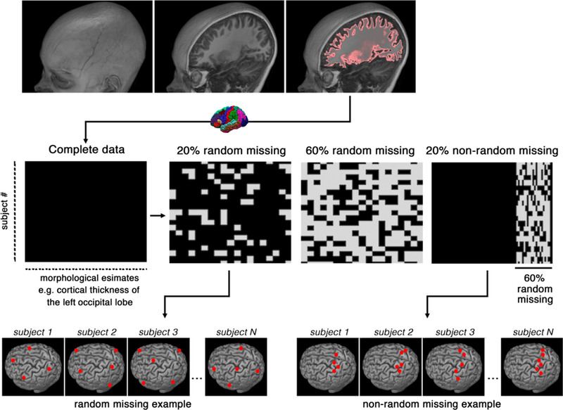
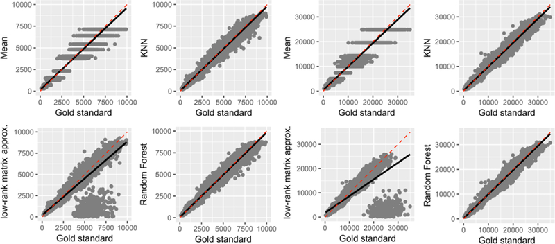
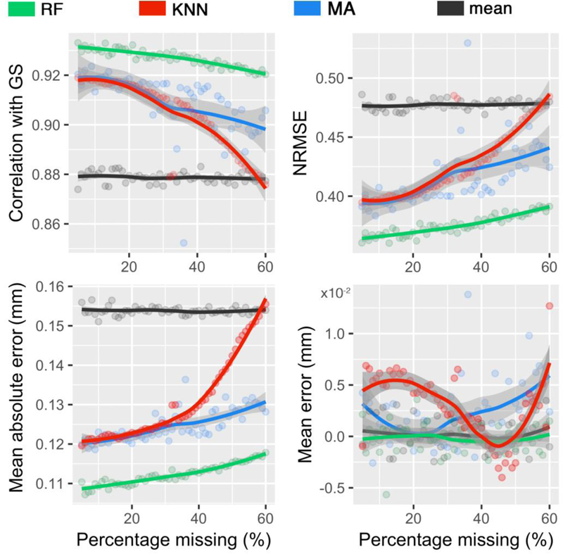
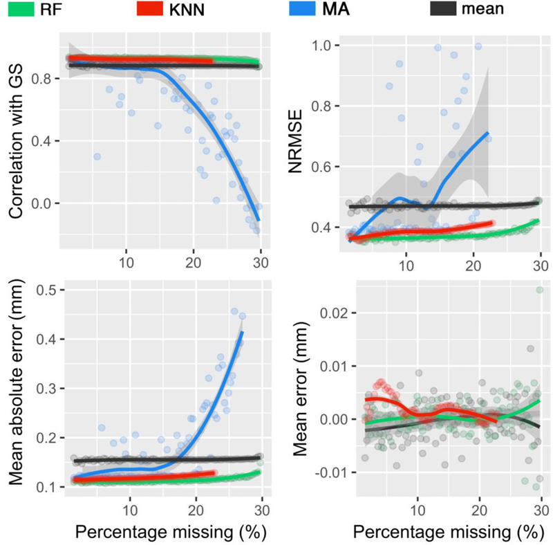
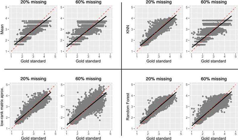
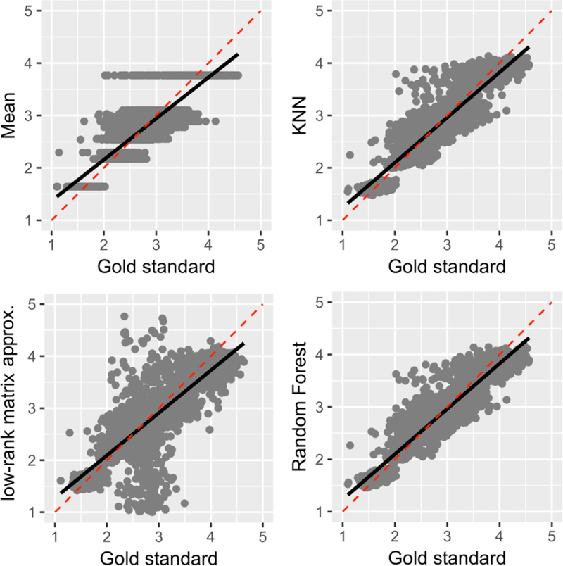
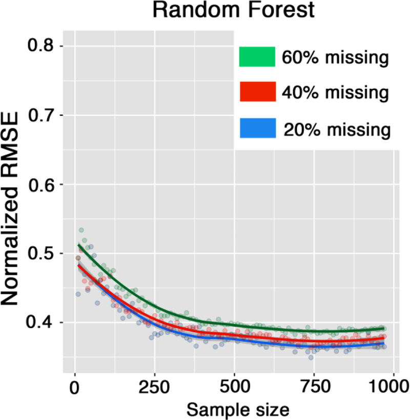
Regional morphological analysis represents a crucial step in most neuroimaging studies. Results from brain segmentation techniques are intrinsically prone to certain degrees of variability, mainly as results of suboptimal segmentation. To reduce this inherent variability, the errors are often identified through visual inspection and then corrected (semi)manually. Identification and correction of incorrect segmentation could be very expensive for large-scale studies. While identification of the incorrect results can be done relatively fast even with manual inspection, the correction step is extremely time-consuming, as it requires training staff to perform laborious manual corrections. Here we frame the correction phase of this problem as a missing data problem. Instead of manually adjusting the segmentation outputs, our computational approach aims to derive accurate morphological measures by machine learning imputation. Data imputation techniques may be used to replace missing or incorrect region average values with carefully chosen imputed values, all of which are computed based on other available multivariate information. We examined our approach of correcting segmentation outputs on a cohort of 970 subjects, which were undergone an extensive, time-consuming, manual post-segmentation correction. A random forest imputation technique recovered the gold standard results with a significant accuracy (r = 0.93, p < 0.0001; when 30% of the segmentations were considered incorrect in a non-random fashion). The random forest technique proved to be most effective for big data studies (N > 250).

摘要

区域形态分析是大多数神经影像学研究的关键步骤。脑分割技术的结果本质上容易受到一定程度的可变性的影响,主要是由于分割不理想。为了减少这种固有变异性,错误通常通过目视检查来识别,然后进行(半)手动校正。对于大规模研究来说,识别和纠正不正确的分割可能非常昂贵。虽然即使通过手动检查也可以相对快速地识别不正确的结果,但校正步骤非常耗时,因为需要培训人员进行费力的手动校正。在这里,我们将该问题的校正阶段框定为缺失数据问题。我们的计算方法不是手动调整分割输出,而是旨在通过机器学习插补来得出准确的形态学度量。数据插补技术可用于用精心选择的插补值替换缺失或不正确的区域平均值,所有这些值都是基于其他可用的多元信息计算得出的。我们在一个由 970 名受试者组成的队列上检查了我们的校正分割输出的方法,这些受试者经历了广泛的、耗时的、手动的分割后校正。随机森林插补技术以显著的准确性(r=0.93,p<0.0001;当以非随机方式考虑 30%的分割不正确时)恢复了金标准结果。随机森林技术对于大数据研究(N>250)最有效。

https://cdn.ncbi.nlm.nih.gov/pmc/blobs/a760/6829024/be275ac21a1f/nihms-1028326-f0007.jpg
https://cdn.ncbi.nlm.nih.gov/pmc/blobs/a760/6829024/9c5362af4b8b/nihms-1028326-f0001.jpg
https://cdn.ncbi.nlm.nih.gov/pmc/blobs/a760/6829024/0c454a7eeab3/nihms-1028326-f0002.jpg
https://cdn.ncbi.nlm.nih.gov/pmc/blobs/a760/6829024/9b8affb5b8fc/nihms-1028326-f0003.jpg
https://cdn.ncbi.nlm.nih.gov/pmc/blobs/a760/6829024/0dae28e46714/nihms-1028326-f0004.jpg
https://cdn.ncbi.nlm.nih.gov/pmc/blobs/a760/6829024/6b0426bd4f49/nihms-1028326-f0005.jpg
https://cdn.ncbi.nlm.nih.gov/pmc/blobs/a760/6829024/ced29719e724/nihms-1028326-f0006.jpg
https://cdn.ncbi.nlm.nih.gov/pmc/blobs/a760/6829024/be275ac21a1f/nihms-1028326-f0007.jpg

相似文献

1
Imputation Strategy for Reliable Regional MRI Morphological Measurements.可靠的区域 MRI 形态学测量的插补策略。
Neuroinformatics. 2020 Jan;18(1):59-70. doi: 10.1007/s12021-019-09426-x.
2
Quality control strategies for brain MRI segmentation and parcellation: Practical approaches and recommendations - insights from the Maastricht study.脑 MRI 分割和分区的质量控制策略:实用方法和建议 - 马斯特里赫特研究的见解。
Neuroimage. 2021 Aug 15;237:118174. doi: 10.1016/j.neuroimage.2021.118174. Epub 2021 May 15.
3
Robust Machine Learning-Based Correction on Automatic Segmentation of the Cerebellum and Brainstem.基于稳健机器学习的小脑和脑干自动分割校正
PLoS One. 2016 May 23;11(5):e0156123. doi: 10.1371/journal.pone.0156123. eCollection 2016.
4
Hippocampus and amygdala volumes from magnetic resonance images in children: Assessing accuracy of FreeSurfer and FSL against manual segmentation.儿童磁共振图像中的海马体和杏仁核体积:评估FreeSurfer和FSL相对于手动分割的准确性。
Neuroimage. 2016 Apr 1;129:1-14. doi: 10.1016/j.neuroimage.2016.01.038. Epub 2016 Jan 26.
5
Multi-atlas segmentation of the whole hippocampus and subfields using multiple automatically generated templates.使用多个自动生成的模板对整个海马体及其子区进行多图谱分割。
Neuroimage. 2014 Nov 1;101:494-512. doi: 10.1016/j.neuroimage.2014.04.054. Epub 2014 Apr 29.
6
Evaluating accuracy of striatal, pallidal, and thalamic segmentation methods: Comparing automated approaches to manual delineation.评估纹状体、苍白球和丘脑分割方法的准确性:比较自动方法与手动勾画。
Neuroimage. 2018 Apr 15;170:182-198. doi: 10.1016/j.neuroimage.2017.02.069. Epub 2017 Mar 1.
7
An efficient and accurate method for robust inter-dataset brain extraction and comparisons with 9 other methods.一种高效准确的稳健跨数据集脑提取方法,并与其他 9 种方法进行比较。
Hum Brain Mapp. 2018 Nov;39(11):4241-4257. doi: 10.1002/hbm.24243. Epub 2018 Jul 4.
8
Using multiple imputation to efficiently correct cerebral MRI whole brain lesion and atrophy data in patients with multiple sclerosis.使用多重填补法有效校正多发性硬化症患者的脑磁共振成像全脑病变和萎缩数据。
Neuroimage. 2015 Oct 1;119:81-8. doi: 10.1016/j.neuroimage.2015.06.037. Epub 2015 Jun 18.
9
Qoala-T: A supervised-learning tool for quality control of FreeSurfer segmented MRI data.Qoala-T:一种用于 FreeSurfer 分割 MRI 数据质量控制的监督学习工具。
Neuroimage. 2019 Apr 1;189:116-129. doi: 10.1016/j.neuroimage.2019.01.014. Epub 2019 Jan 8.
10
Machine learning identifies stroke features between species.机器学习识别物种间的中风特征。
Theranostics. 2021 Jan 1;11(6):3017-3034. doi: 10.7150/thno.51887. eCollection 2021.

引用本文的文献

1
Socioeconomic status, prefrontal cortical volume, and cardiometabolic risk in early adolescence.青少年早期的社会经济地位、前额叶皮质体积与心脏代谢风险
Health Psychol. 2025 May 5. doi: 10.1037/hea0001508.
2
Dementia-related user-based collaborative filtering for imputing missing data and generating a reliability scale on clinical test scores.基于用户的与痴呆症相关的协同过滤,用于填补缺失数据和生成临床测试分数的可靠性量表。
PeerJ. 2022 May 26;10:e13425. doi: 10.7717/peerj.13425. eCollection 2022.
3
Three-dimensional self-attention conditional GAN with spectral normalization for multimodal neuroimaging synthesis.

本文引用的文献

1
Qoala-T: A supervised-learning tool for quality control of FreeSurfer segmented MRI data.Qoala-T:一种用于 FreeSurfer 分割 MRI 数据质量控制的监督学习工具。
Neuroimage. 2019 Apr 1;189:116-129. doi: 10.1016/j.neuroimage.2019.01.014. Epub 2019 Jan 8.
2
Identifying errors in Freesurfer automated skull stripping and the incremental utility of manual intervention.识别 Freesurfer 自动颅骨剥离中的错误和手动干预的增量效用。
Brain Imaging Behav. 2019 Oct;13(5):1281-1291. doi: 10.1007/s11682-018-9951-8.
3
Neuroanatomical morphometric characterization of sex differences in youth using statistical learning.
基于谱归一化的三维自注意力条件生成对抗网络在多模态神经影像合成中的应用。
Magn Reson Med. 2021 Sep;86(3):1718-1733. doi: 10.1002/mrm.28819. Epub 2021 May 7.
4
Global and Regional Changes in Perivascular Space in Idiopathic and Familial Parkinson's Disease.特发性和家族性帕金森病患者的血管周围间隙的全球和区域变化。
Mov Disord. 2021 May;36(5):1126-1136. doi: 10.1002/mds.28473. Epub 2021 Jan 20.
5
Volumetric distribution of perivascular space in relation to mild cognitive impairment.血管周围间隙容积分布与轻度认知障碍的关系。
Neurobiol Aging. 2021 Mar;99:28-43. doi: 10.1016/j.neurobiolaging.2020.12.010. Epub 2020 Dec 16.
6
Investigating the Added Value of FreeSurfer's Manual Editing Procedure for the Study of the Reading Network in a Pediatric Population.探究FreeSurfer手动编辑程序在儿科人群阅读网络研究中的附加价值。
Front Hum Neurosci. 2020 Apr 24;14:143. doi: 10.3389/fnhum.2020.00143. eCollection 2020.
利用统计学习方法对青少年性别差异的神经解剖形态进行特征描述。
Neuroimage. 2018 May 15;172:217-227. doi: 10.1016/j.neuroimage.2018.01.065. Epub 2018 Feb 3.
4
Cortical thickness alterations linked to somatoform and psychological dissociation in functional neurological disorders.与功能性神经障碍中躯体形式和心理解离相关的皮质厚度改变。
Hum Brain Mapp. 2018 Jan;39(1):428-439. doi: 10.1002/hbm.23853. Epub 2017 Oct 28.
5
Comparison of accuracy between FSL's FIRST and Freesurfer for caudate nucleus and putamen segmentation.比较 FSL 的 FIRST 和 Freesurfer 在尾状核和壳核分割上的准确性。
Sci Rep. 2017 May 25;7(1):2418. doi: 10.1038/s41598-017-02584-5.
6
Evaluating accuracy of striatal, pallidal, and thalamic segmentation methods: Comparing automated approaches to manual delineation.评估纹状体、苍白球和丘脑分割方法的准确性:比较自动方法与手动勾画。
Neuroimage. 2018 Apr 15;170:182-198. doi: 10.1016/j.neuroimage.2017.02.069. Epub 2017 Mar 1.
7
Brain development during adolescence: A mixed-longitudinal investigation of cortical thickness, surface area, and volume.青少年时期的大脑发育:皮质厚度、表面积和体积的混合纵向研究。
Hum Brain Mapp. 2016 Jun;37(6):2027-38. doi: 10.1002/hbm.23154. Epub 2016 Mar 4.
8
Structural Neuroimaging Genetics Interactions in Alzheimer's Disease.阿尔茨海默病中的结构神经影像遗传学相互作用
J Alzheimers Dis. 2015;48(4):1051-63. doi: 10.3233/JAD-150335.
9
Big biomedical data as the key resource for discovery science.大生物医学数据作为发现科学的关键资源。
J Am Med Inform Assoc. 2015 Nov;22(6):1126-31. doi: 10.1093/jamia/ocv077. Epub 2015 Jul 21.
10
The Philadelphia Neurodevelopmental Cohort: A publicly available resource for the study of normal and abnormal brain development in youth.费城神经发育队列研究:一个可公开获取的资源,用于研究青少年正常和异常的大脑发育。
Neuroimage. 2016 Jan 1;124(Pt B):1115-1119. doi: 10.1016/j.neuroimage.2015.03.056. Epub 2015 Mar 31.