Department of Neurology and Neurological Sciences, Stanford University, 300 Pasteur Dr. Room H3144, MC 5235, Stanford, CA 94305, United States of America.
Department of Radiology, Stanford University, 1201 Welch Road. Room PS-064, MC 5488, Stanford, CA 94305, United States of America.
Neuroimage Clin. 2019;23:101824. doi: 10.1016/j.nicl.2019.101824. Epub 2019 Apr 18.
Parkinson's disease (PD) episodic memory impairments are common; however, it is not known whether these impairments are due to hippocampal pathology. Hippocampal Lewy-bodies emerge by Braak stage 4, but are not uniformly distributed. For instance, hippocampal CA1 Lewy-body pathology has been specifically associated with pre-mortem episodic memory performance in demented patients. By contrast, the dentate gyrus (DG) is relatively free of Lewy-body pathology. In this study, we used ultra-high field 7-Tesla to measure hippocampal subfields in vivo and determine if these measures predict episodic memory impairment in PD during life.
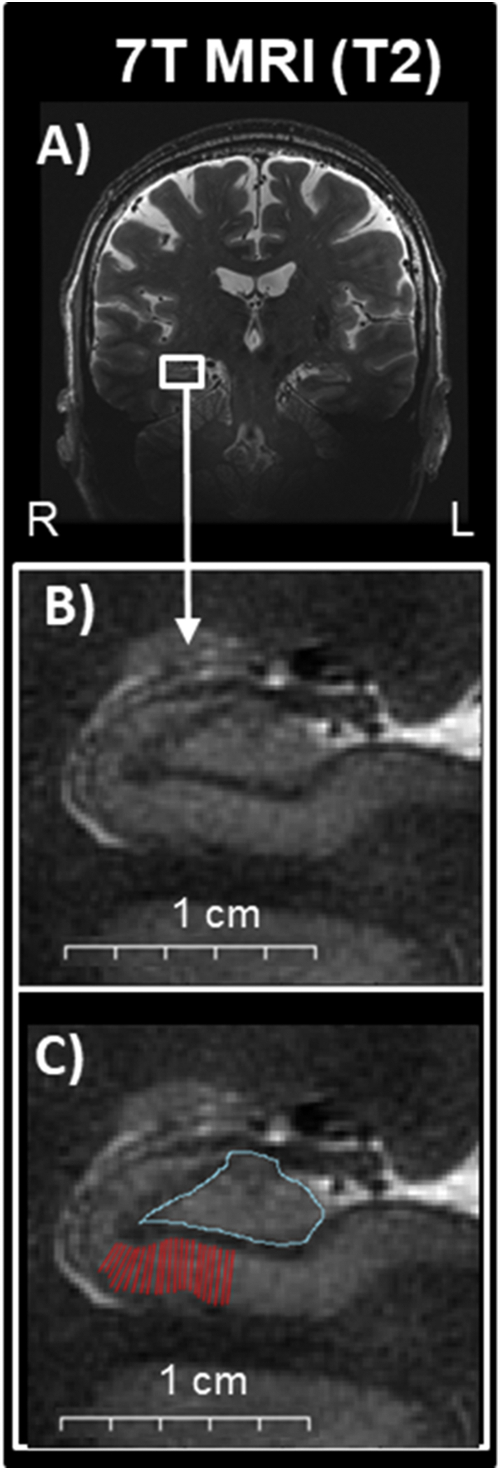
We studied 29 participants with PD (age 65.5 ± 7.8 years; disease duration 4.5 ± 3.0 years) and 8 matched-healthy controls (age 67.9 ± 6.8 years), who completed comprehensive neuropsychological testing and MRI. With 7-Tesla MRI, we used validated segmentation techniques to estimate CA1 stratum pyramidale (CA1-SP) and stratum radiatum lacunosum moleculare (CA1-SRLM) thickness, dentate gyrus/CA3 (DG/CA3) area, and whole hippocampus area. We used linear regression, which included imaging and clinical measures (age, duration, education, gender, and CSF), to determine the best predictors of episodic memory impairment in PD.
In our cohort, 62.1% of participants with PD had normal cognition, 27.6% had mild cognitive impairment, and 10.3% had dementia. Using 7-Tesla MRI, we found that smaller CA1-SP thickness was significantly associated with poorer immediate memory, delayed memory, and delayed cued memory; by contrast, whole hippocampus area, DG/CA3 area, and CA1-SRLM thickness did not significantly predict memory. Age-adjusted linear regression models revealed that CA1-SP predicted immediate memory (beta[standard error]10.895[4.215], p < .05), delayed memory (12.740[5.014], p < .05), and delayed cued memory (12.801[3.991], p < .05). In the fully-adjusted models, which included all five clinical measures as covariates, only CA1-SP remained a significant predictor of delayed cued memory (13.436[4.651], p < .05).
In PD, we found hippocampal CA1-SP subfield thickness estimated on 7-Tesla MRI scans was the best predictor of episodic memory impairment, even when controlling for confounding clinical measures. Our results imply that ultra-high field imaging could be a sensitive measure to identify changes in hippocampal subfields and thus probe the neuroanatomical underpinnings of episodic memory impairments in patients with PD.
帕金森病(PD)的情景记忆损伤很常见;然而,这些损伤是否是由于海马体病理学引起的尚不清楚。海马Lewy 体出现在 Braak 阶段 4,但分布不均匀。例如,海马 CA1 Lewy 体病理学与痴呆患者生前的情景记忆表现有具体关联。相比之下,齿状回(DG)相对没有 Lewy 体病理学。在这项研究中,我们使用超高场 7-Tesla 测量体内海马亚区,并确定这些指标是否可以预测 PD 患者在有生之年的情景记忆损伤。
我们研究了 29 名 PD 患者(年龄 65.5±7.8 岁;疾病持续时间 4.5±3.0 年)和 8 名匹配的健康对照组(年龄 67.9±6.8 岁),他们完成了全面的神经心理学测试和 MRI。使用 7-Tesla MRI,我们使用经过验证的分割技术来估计 CA1 锥体层(CA1-SP)和放射层(CA1-SRLM)厚度、齿状回/CA3(DG/CA3)区域和整个海马区面积。我们使用线性回归,其中包括影像学和临床指标(年龄、持续时间、教育、性别和 CSF),以确定 PD 患者情景记忆损伤的最佳预测指标。
在我们的队列中,62.1%的 PD 患者认知正常,27.6%的患者有轻度认知障碍,10.3%的患者有痴呆症。使用 7-Tesla MRI,我们发现较小的 CA1-SP 厚度与较差的即时记忆、延迟记忆和延迟提示记忆显著相关;相比之下,整个海马区面积、DG/CA3 面积和 CA1-SRLM 厚度与记忆无显著相关性。年龄调整的线性回归模型显示 CA1-SP 预测即时记忆(β[标准误差]10.895[4.215],p<0.05)、延迟记忆(12.740[5.014],p<0.05)和延迟提示记忆(12.801[3.991],p<0.05)。在包含所有五个临床指标作为协变量的完全调整模型中,只有 CA1-SP 仍然是延迟提示记忆的显著预测指标(13.436[4.651],p<0.05)。
在 PD 中,我们发现 7-Tesla MRI 扫描估计的海马 CA1-SP 亚区厚度是情景记忆损伤的最佳预测指标,即使在控制混杂的临床指标后也是如此。我们的结果表明,超高场成像可能是一种敏感的测量方法,可以识别海马亚区的变化,并由此探究 PD 患者情景记忆损伤的神经解剖学基础。