Bonilla Ximena, Vanegas Natalia-Del Pilar, Vernot Jean Paul
Cellular and Molecular Physiology, Faculty of Medicine, Universidad Nacional de Colombia, Bogotá D.C. 111321, Colombia.
Biomedical Research Institute, Faculty of Medicine, Universidad Nacional de Colombia, Bogotá D.C. 111321, Colombia.
Stem Cells Int. 2019 Apr 1;2019:3864948. doi: 10.1155/2019/3864948. eCollection 2019.
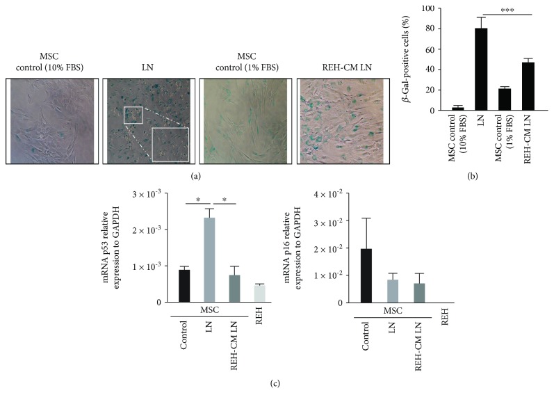
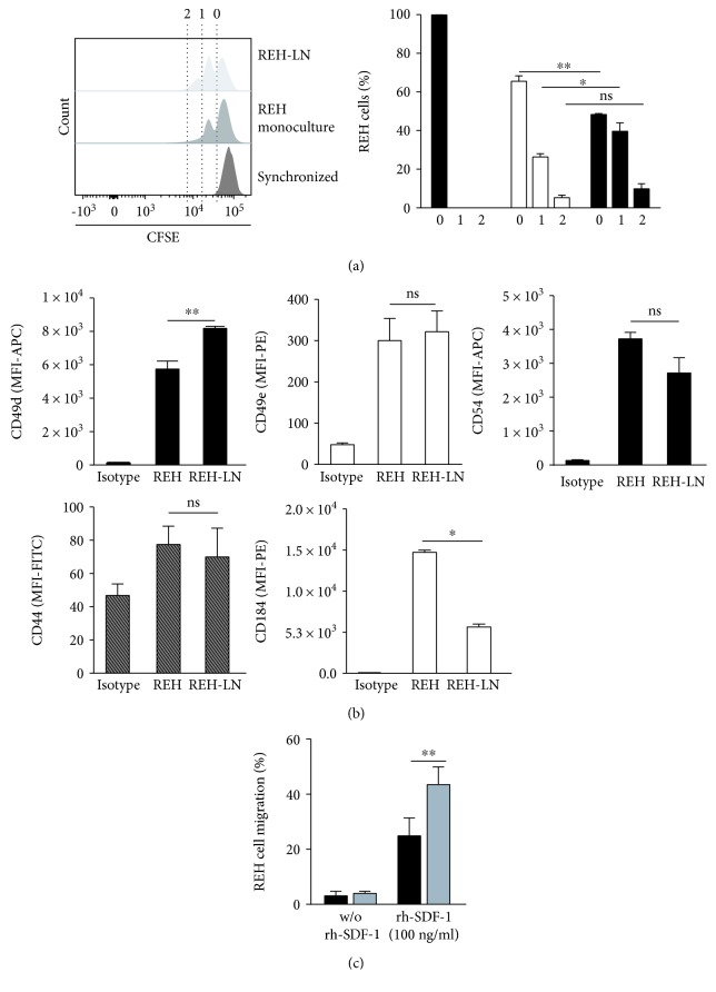
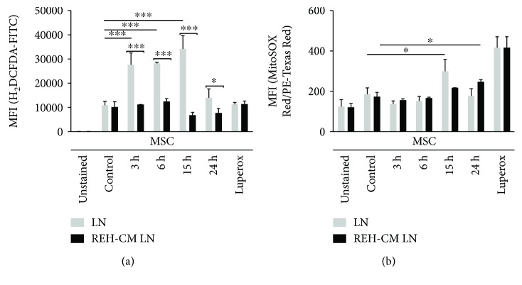
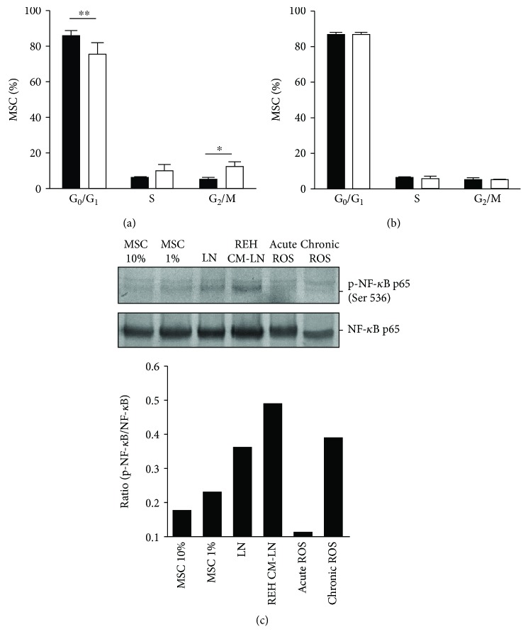
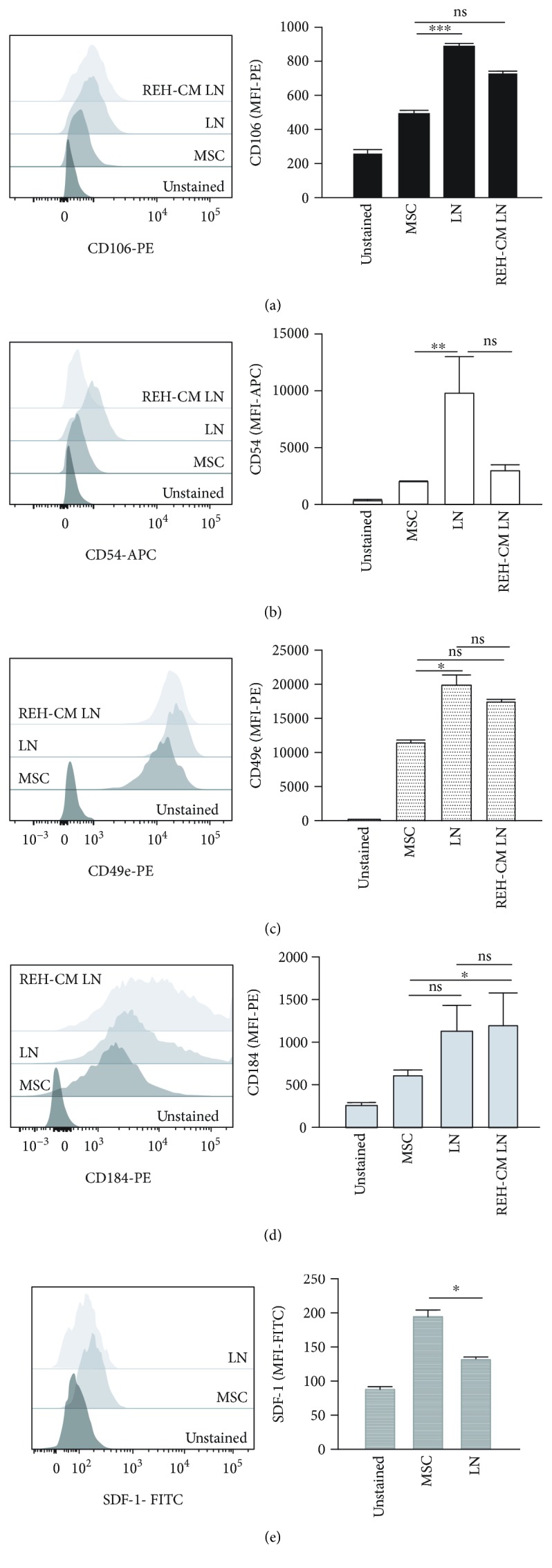
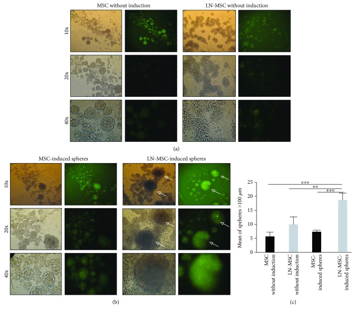
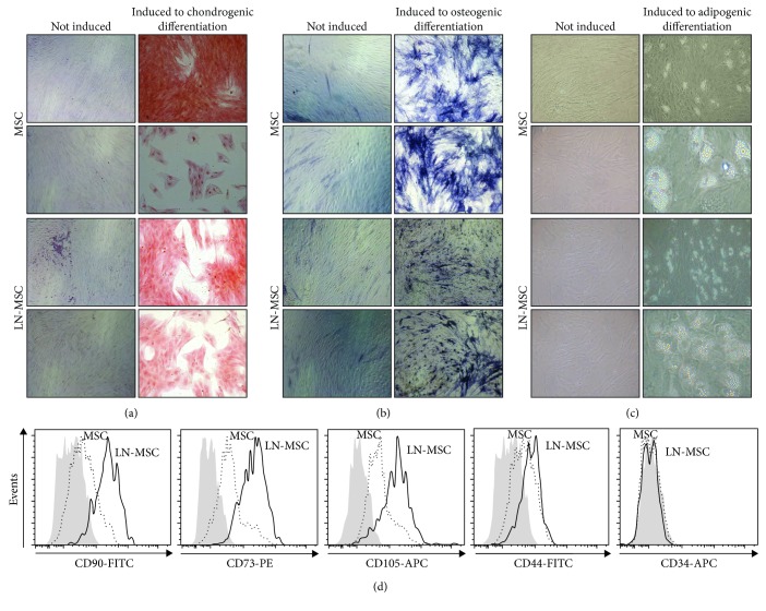
Mesenchymal stem cells (MSC) constitute an important cell population of the bone marrow hematopoietic niche that supports normally hematopoietic stem cells (HSC) but eventually also leukemic cells. The alterations that occur in the MSC under leukemic stress are not well known. To deepen on this topic, we have used an model of the leukemic niche (LN) by coculturing MSC with an acute lymphocytic leukemia cell line (REH) and proceeded to evaluate MSC characteristics and functions. We found that leukemic cells induced in MSC a significant increase both in senescence-associated -galactosidase activity and in p53 gene expression. MSC in the LN also showed a persistent production of cytoplasmic reactive oxygen species (ROS) and a G2/M phase arrest of the cell cycle. Another acute leukemic cell line (SUP-B15) produced almost the same effects on MSC. REH cells adhere strongly to MSC possibly as a result of an increased expression of the adhesion molecules VCAM-1, ICAM-1, and CD49e in MSC and of CD49d in REH cells. Although mesensphere formation was normal or even increased, multipotent differentiation capacity was impaired in MSC from the LN. A REH-conditioned medium was only partially (about 50%) capable of inducing the same changes in MSC, suggesting that cell-to-cell contact is more efficient in inducing these changes. Despite these important effects on MSC in the LN, REH cells increased their cell adhesion, proliferation rate, and directed-migration capacity. In conclusion, in this LN model, leukemic cells affect importantly the MSC, inducing a senescence process that seems to favour leukemic cell growth.
间充质干细胞(MSC)是骨髓造血微环境中的重要细胞群体,它通常支持造血干细胞(HSC),但最终也支持白血病细胞。白血病应激下MSC发生的改变尚不清楚。为了深入研究这个问题,我们通过将MSC与急性淋巴细胞白血病细胞系(REH)共培养建立了白血病微环境(LN)模型,并对MSC的特性和功能进行了评估。我们发现白血病细胞可诱导MSC中衰老相关β-半乳糖苷酶活性和p53基因表达显著增加。LN中的MSC还表现出持续产生细胞质活性氧(ROS)以及细胞周期的G2/M期阻滞。另一种急性白血病细胞系(SUP-B15)对MSC产生了几乎相同的影响。REH细胞与MSC强烈黏附,这可能是由于MSC中黏附分子VCAM-1、ICAM-1和CD49e以及REH细胞中CD49d表达增加所致。尽管球状体形成正常甚至增加,但LN中MSC的多能分化能力受损。REH条件培养基仅部分(约50%)能够诱导MSC发生相同的变化,这表明细胞间接触在诱导这些变化方面更有效。尽管对LN中的MSC有这些重要影响,但REH细胞增加了其细胞黏附、增殖率和定向迁移能力。总之,在这个LN模型中,白血病细胞对MSC有重要影响,诱导了一个似乎有利于白血病细胞生长的衰老过程。