Neurotrauma and Ophthalmology Research Group, School of Clinical and Experimental Medicine, College of Medical and Dental Sciences, University of Birmingham, Edgbaston, Birmingham B15 2TT, UK.
National Institute for Health Research Surgical Reconstruction and Microbiology Research Centre, Queen Elizabeth Hospital, Edgbaston, Birmingham B15 2TH, UK.
Int J Mol Sci. 2019 May 7;20(9):2239. doi: 10.3390/ijms20092239.
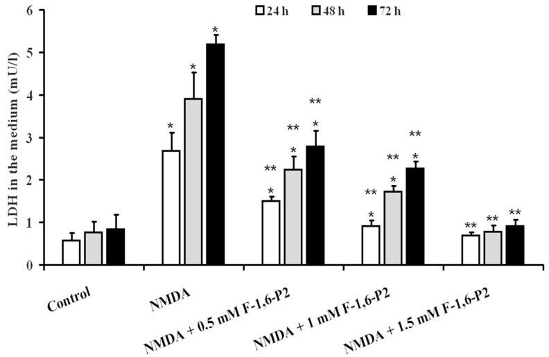
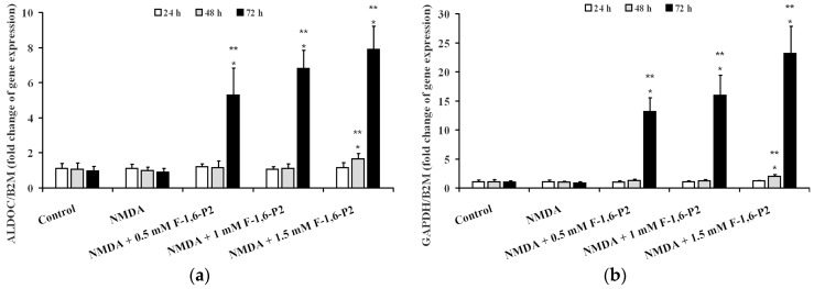
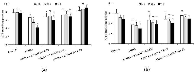
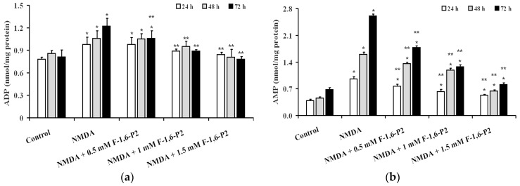
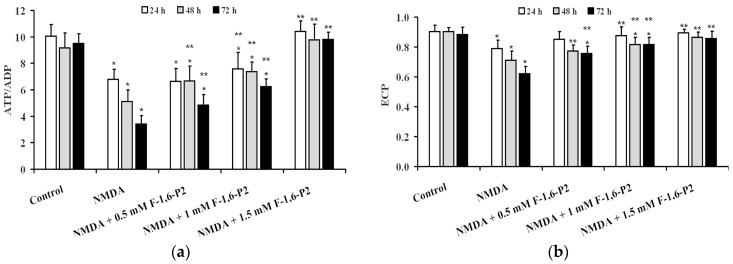
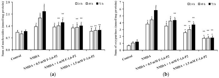
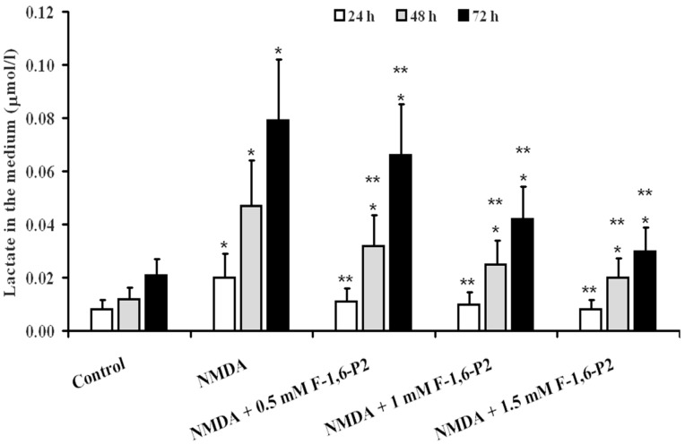
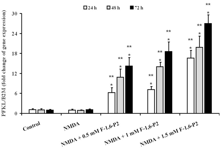
Effects of fructose 1,6-bisphosphate (F-1,6-P2) towards -methyl-d-aspartate NMDA excitotoxicity were evaluated in rat organotypic hippocampal brain slice cultures (OHSC) challenged for 3 h with 30 μM NMDA, followed by incubations (24, 48, and 72 h) without (controls) and with F-1,6-P2 (0.5, 1 or 1.5 mM). At each time, cell necrosis was determined by measuring LDH in the medium. Energy metabolism was evaluated by measuring ATP, GTP, ADP, AMP, and ATP catabolites (nucleosides and oxypurines) in deproteinized OHSC extracts. Gene expressions of phosphofructokinase, aldolase, and glyceraldehyde-3-phosphate dehydrogenase were also measured. F-1,6-P2 dose-dependently decreased NMDA excitotoxicity, abolishing cell necrosis at the highest concentration tested (1.5 mM). Additionally, F-1,6-P2 attenuated cell energy imbalance caused by NMDA, ameliorating the mitochondrial phosphorylating capacity (increase in ATP/ADP ratio) Metabolism normalization occurred when using 1.5 mM F-1,6-P2. Remarkable increase in expressions of phosphofructokinase, aldolase and glyceraldehyde-3-phosphate dehydrogenase (up to 25 times over the values of controls) was also observed. Since this phenomenon was recorded even in OHSC treated with F-1,6-P2 with no prior challenge with NMDA, it is highly conceivable that F-1,6-P2 can enter into intact cerebral cells producing significant benefits on energy metabolism. These effects are possibly mediated by changes occurring at the gene level, thus opening new perspectives for F-1,6-P2 application as a useful adjuvant to rescue mitochondrial metabolism of cerebral cells under stressing conditions.
1,6-二磷酸果糖(F-1,6-P2)对 30μM NMDA 诱导的大鼠器官型海马脑片(OHSC)培养物兴奋性毒性的影响,在 3 h 内用 30μM NMDA 进行挑战,随后在没有(对照组)和有 F-1,6-P2(0.5、1 或 1.5 mM)的情况下孵育(24、48 和 72 h)。在每个时间点,通过测量培养基中的 LDH 来确定细胞坏死。通过测量去蛋白 OHSC 提取物中的 ATP、GTP、ADP、AMP 和 ATP 代谢物(核苷和氧嘌呤)来评估能量代谢。还测量了磷酸果糖激酶、醛缩酶和甘油醛-3-磷酸脱氢酶的基因表达。F-1,6-P2 剂量依赖性地降低了 NMDA 兴奋性毒性,在测试的最高浓度(1.5 mM)时消除了细胞坏死。此外,F-1,6-P2 减轻了 NMDA 引起的细胞能量失衡,改善了线粒体磷酸化能力(增加 ATP/ADP 比)。当使用 1.5 mM F-1,6-P2 时,代谢恢复正常。还观察到磷酸果糖激酶、醛缩酶和甘油醛-3-磷酸脱氢酶的表达显著增加(比对照组增加 25 倍)。由于即使在没有预先用 NMDA 处理的 OHSC 中也记录到这种现象,因此可以高度推测 F-1,6-P2 可以进入完整的脑细胞,对能量代谢产生显著益处。这些效应可能是通过基因水平的变化介导的,从而为 F-1,6-P2 的应用开辟了新的前景,作为一种有用的辅助手段,在应激条件下挽救脑细胞的线粒体代谢。