Jang Sooah, Kim Hyunjeong, Kim Hye-Jin, Lee Su Kyoung, Kim Eun Woo, Namkoong Kee, Kim Eosu
Department of Psychiatry, Yonsei University College of Medicine, Seoul, Republic of Korea.
Institute of Behavioral Science in Medicine, Yonsei University College of Medicine, Seoul, Republic of Korea.
Psychiatry Investig. 2018 Feb;15(2):205-213. doi: 10.30773/pi.2017.04.02. Epub 2017 Nov 29.
Conventional methods for organotypic hippocampal tissue slice culture (OHSC) have shown several disadvantages or limitations regarding age of animals used, duration of culture and difficulty using neurodegenerative models. Therefore, we tried to establish OHSC from old 3xTg-Alzheimer's disease (AD) mice for longer period (over 4 weeks) and to validate utility of this system as a valid platform for translational neuroscience of AD.
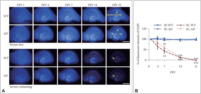
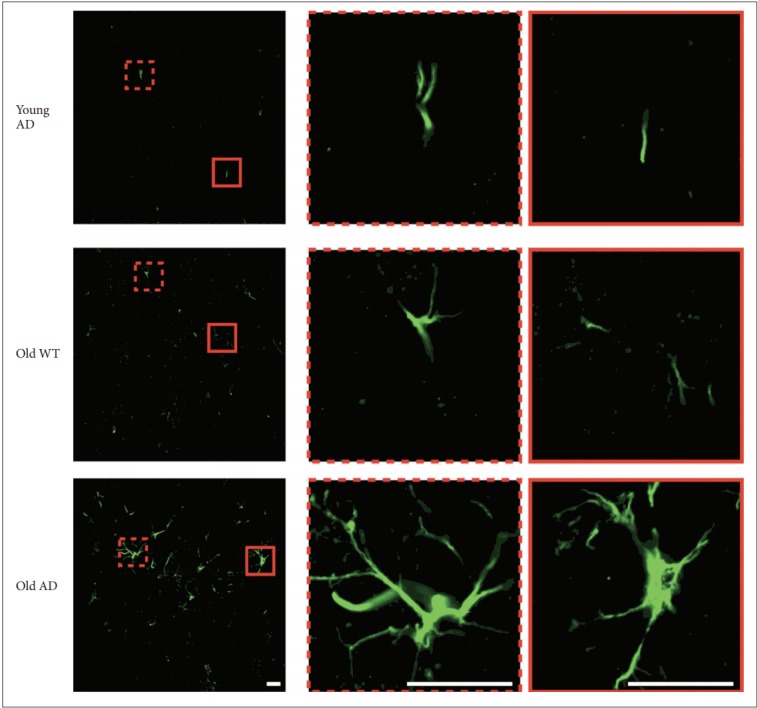
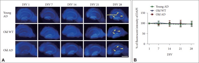
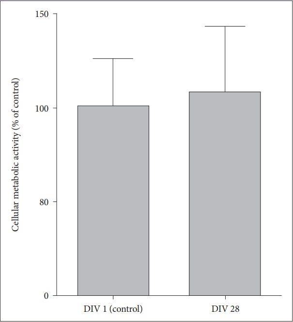
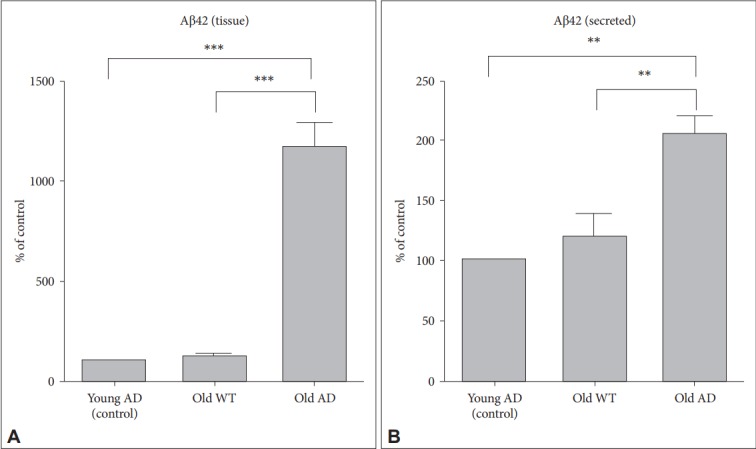
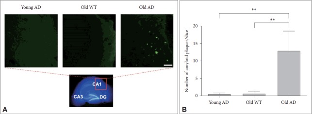
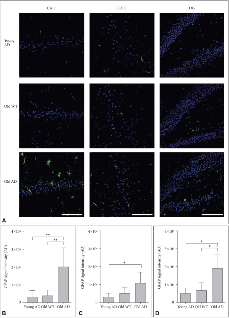
OHSC was performed with old 3xTg-AD mice (12-14 months), old wild type mice (12-14 months) and young 3xTg-AD mice (2-4 months) using serum-free medium for 4 weeks. Hippocampal structure was evaluated by 4', 6-diamidino-2-phenylindole (DAPI) intensity and neuronal metabolism was measured by Alamarblue assay. Pathologic characteristics of AD were also investigated; β-amyloid levels by ELISA, amyloid plaque deposition by Thioflavin-S staining, and glial activation by immunohistochemistry.
Following 4-week culture in serum-free media, hippocampal cells and layers were well preserved in cultured slices from old AD mice as was in those from young AD and old wild type mice. On the contrary, excessive regression of total visible cells was observed in conventional serum-containing medium regardless of genotype of mice. In parallel with this well preserved structure, major pathologic characteristics of AD were also well manifested in hippocampal slices from old AD mice.
Our findings suggest that long-term OHSC from old 3xTg-AD mouse can serve as a promising system for studies on pathophysiology of AD, especially with the minimum number of sacrifice of experimental animals.
传统的器官型海马组织切片培养(OHSC)方法在所用动物年龄、培养持续时间以及使用神经退行性疾病模型的难度方面存在若干缺点或局限性。因此,我们试图从老年3xTg-阿尔茨海默病(AD)小鼠建立OHSC并进行更长时间(超过4周)的培养,以验证该系统作为AD转化神经科学有效平台的实用性。
使用无血清培养基对老年3xTg-AD小鼠(12 - 14个月)、老年野生型小鼠(12 - 14个月)和年轻3xTg-AD小鼠(2 - 4个月)进行OHSC培养4周。通过4',6-二脒基-2-苯基吲哚(DAPI)强度评估海马结构,通过Alamarblue检测法测量神经元代谢。还研究了AD的病理特征;通过酶联免疫吸附测定(ELISA)检测β-淀粉样蛋白水平,通过硫黄素-S染色检测淀粉样斑块沉积,通过免疫组织化学检测胶质细胞活化。
在无血清培养基中培养4周后,老年AD小鼠培养切片中的海马细胞和层与年轻AD小鼠和老年野生型小鼠的培养切片一样保存良好。相反,无论小鼠基因型如何,在传统的含血清培养基中均观察到总可见细胞过度退化。与这种保存良好的结构并行,老年AD小鼠海马切片中AD的主要病理特征也得到了很好的体现。
我们的研究结果表明,来自老年3xTg-AD小鼠的长期OHSC可作为研究AD病理生理学的一个有前景的系统,特别是在实验动物牺牲数量最少的情况下。