Memory and Aging Center, University of California San Francisco, San Francisco, CA 94143, USA; Helen Wills Neuroscience Institute, University of California Berkeley, Berkeley, CA 94720, USA; Department of Neurology & Alzheimer Center, Neuroscience Campus Amsterdam, VU University Medical Center, Amsterdam 1081 HZ, the Netherlands.
Memory and Aging Center, University of California San Francisco, San Francisco, CA 94143, USA.
Neuroimage Clin. 2019;23:101848. doi: 10.1016/j.nicl.2019.101848. Epub 2019 May 2.
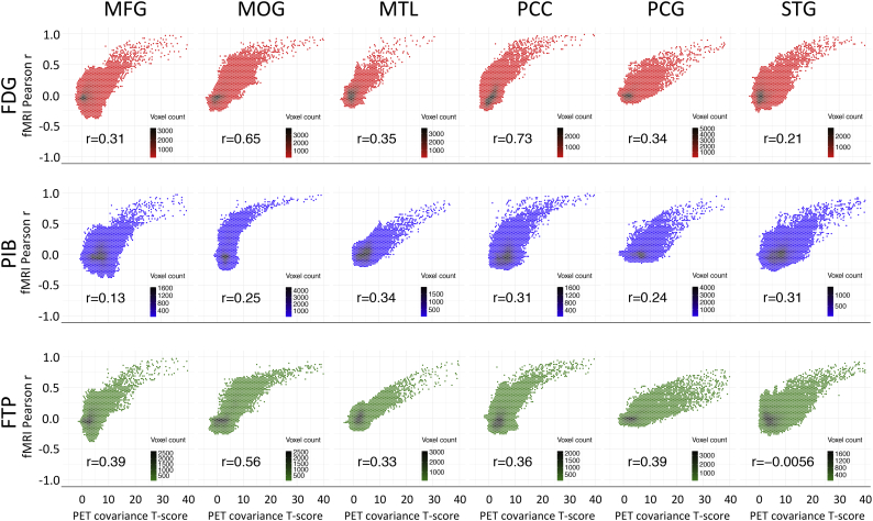
According to the network model of neurodegeneration, the spread of pathogenic proteins occurs selectively along connected brain regions. We tested in vivo whether the distribution of filamentous tau (measured with [F]flortaucipir-PET), fibrillar amyloid-β ([C]PIB-PET) and glucose hypometabolism ([F]FDG-PET) follows the intrinsic functional organization of the healthy brain. We included 63 patients with Alzheimer's disease (AD; 30 male, 63 ± 8 years) who underwent [F]flortaucipir, [C]PIB and [F]FDG PET, and 1000 young adults (427 male, 21 ± 3 years) who underwent task-free fMRI. We selected six predefined disease epicenters as seeds for whole-brain voxelwise covariance analyses to compare correlated patterns of tracer uptake across AD patients against fMRI intrinsic connectivity patterns in young adults. We found a striking convergence between [F]flortaucipir covariance patterns and intrinsic connectivity maps (range Spearman rho's: 0.32-0.78, p < .001), which corresponded with expected functional networks (range goodness-of-fit: 3.8-8.2). The topography of amyloid-β covariance patterns was more diffuse and less network-specific, while glucose hypometabolic patterns were more spatially restricted than tau but overlapped with functional networks. These findings suggest that the spatial patterns of tau and glucose hypometabolism observed in AD resemble the functional organization of the healthy brain, supporting the notion that tau pathology spreads through circumscribed brain networks and drives neurodegeneration.
根据神经退行性变的网络模型,致病蛋白的传播是沿着连接的大脑区域选择性发生的。我们在体内测试了丝状 tau(用 [F]flortaucipir-PET 测量)、纤维状淀粉样蛋白-β ([C]PIB-PET) 和葡萄糖代谢低下 ([F]FDG-PET) 的分布是否遵循健康大脑的固有功能组织。我们纳入了 63 名阿尔茨海默病 (AD) 患者(30 名男性,63±8 岁),他们接受了 [F]flortaucipir、[C]PIB 和 [F]FDG PET 检查,以及 1000 名年轻成年人(427 名男性,21±3 岁)接受了无任务 fMRI 检查。我们选择了六个预先定义的疾病中心作为种子,用于全脑体素协方差分析,以比较 AD 患者的示踪剂摄取相关模式与年轻成年人 fMRI 固有连通性模式。我们发现 [F]flortaucipir 协方差模式与固有连通性图谱之间存在惊人的一致性(范围 Spearman rho:0.32-0.78,p<0.001),这与预期的功能网络相对应(范围拟合度:3.8-8.2)。淀粉样蛋白-β协方差模式的拓扑结构更加弥散,与网络的特异性较低,而葡萄糖代谢低下的模式比 tau 更受限制,但与功能网络重叠。这些发现表明,AD 中观察到的 tau 和葡萄糖代谢低下的空间模式类似于健康大脑的功能组织,支持 tau 病理学通过限定的大脑网络传播并驱动神经退行性变的观点。