From the Northwestern University Feinberg Medical School, Chicago, IL (J.T., J.W., M.Y., J.R., M.S., D.B.).
Charité-Universitätsmedizin, Berlin, Germany (J.T., J.R., M.S., M.B.).
Hypertension. 2019 Jul;74(1):83-94. doi: 10.1161/HYPERTENSIONAHA.119.12873. Epub 2019 May 13.
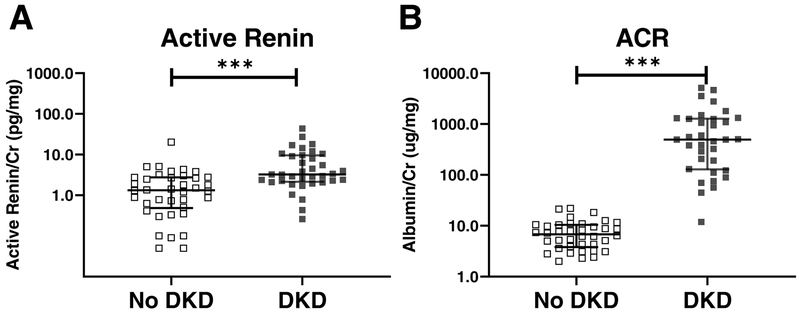
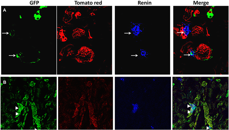
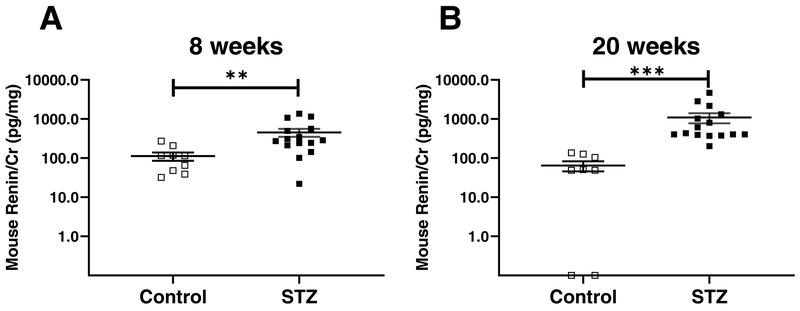
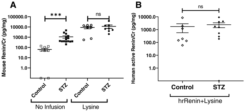
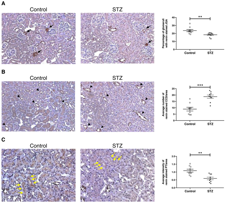
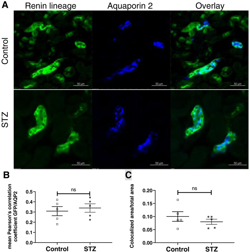
In patients with diabetic kidney disease (DKD), plasma renin activity is usually decreased, but there is limited information on urinary renin and its origin. Urinary renin was evaluated in samples from patients with longstanding type I diabetes mellitus and mice with streptozotocin-induced diabetes mellitus. Renin-reporter mouse model (Ren1d-Cre;mT/mG) was made diabetic with streptozotocin to examine whether the distribution of cells of the renin lineage was altered in a chronic diabetic environment. Active renin was increased in urine samples from patients with DKD (n=36), compared with those without DKD (n=38; 3.2 versus 1.3 pg/mg creatinine; P<0.001). In mice with streptozotocin-induced diabetes mellitus, urine renin was also increased compared with nondiabetic controls. By immunohistochemistry, in mice with streptozotocin-induced diabetes mellitus, juxtaglomerular apparatus and proximal tubular renin staining were reduced, whereas collecting tubule staining, by contrast, was increased. To examine the role of filtration and tubular reabsorption on urinary renin, mice were either infused with either mouse or human recombinant renin and lysine (a blocker of proximal tubular protein reabsorption). Infusion of either form of renin together with lysine markedly increased urinary renin such that it was no longer different between nondiabetic and diabetic mice. Megalin mRNA was reduced in the kidney cortex of streptozotocin-treated mice (0.70±0.09 versus 1.01±0.04 in controls, P=0.01) consistent with impaired tubular reabsorption. In Ren1d-Cre;mT/mG with streptozotocin-induced diabetes mellitus, the distribution of renin lineage cells within the kidney was similar to nondiabetic renin-reporter mice. No evidence for migration of cells of renin linage to the collecting duct in diabetic mice could be found. Renin mRNA in microdissected collecting ducts from streptozotocin-treated mice, moreover, was not significantly different than in controls, whereas in kidney cortex, largely reflecting juxtaglomerular apparatus renin, it was significantly reduced. In conclusion, in urine from patients with type 1 diabetes mellitus and DKD and from mice with streptozotocin-induced diabetes mellitus, renin is elevated. This cannot be attributed to production from cells of the renin lineage migrating to the collecting duct in a chronic hyperglycemic environment. Rather, the elevated levels of urinary renin found in DKD are best attributed to altered glomerular filteration and impaired proximal tubular reabsorption.
在患有糖尿病肾病(DKD)的患者中,血浆肾素活性通常降低,但有关尿肾素及其来源的信息有限。在患有长期 1 型糖尿病的患者和链脲佐菌素诱导的糖尿病小鼠的样本中评估了肾素。使用链脲佐菌素使 Ren1d-Cre;mT/mG 肾素报告小鼠模型发生糖尿病,以检查在慢性糖尿病环境中肾素谱系细胞的分布是否发生改变。与无 DKD 的患者(36 名,3.2pg/mg 肌酐;P<0.001)相比,DKD 患者的尿肾素增加(36 名,3.2pg/mg 肌酐;P<0.001)。与非糖尿病对照组相比,链脲佐菌素诱导的糖尿病小鼠的尿肾素也增加。通过免疫组织化学,在链脲佐菌素诱导的糖尿病小鼠中,肾小球旁器和近端肾小管肾素染色减少,而集合管染色增加。为了检查滤过和肾小管重吸收对尿肾素的作用,用鼠或人重组肾素和赖氨酸(近端肾小管蛋白重吸收的阻滞剂)分别对小鼠进行输注。与非糖尿病小鼠相比,输注任何形式的肾素加赖氨酸均显着增加尿肾素,使其不再不同。链脲佐菌素处理的小鼠肾皮质中的 megalin mRNA 减少(0.70±0.09 与对照组的 1.01±0.04,P=0.01),与肾小管重吸收受损一致。在 Ren1d-Cre;mT/mG 与链脲佐菌素诱导的糖尿病中,肾脏中肾素谱系细胞的分布与非糖尿病肾素报告小鼠相似。在糖尿病小鼠中未发现肾素谱系细胞向集合管迁移的证据。此外,与对照组相比,从小鼠微分离的集合管中提取的肾素 mRNA 没有显着差异,而在肾皮质中,主要反映肾小球旁器肾素,其水平显着降低。总之,在患有 1 型糖尿病和 DKD 的患者的尿液以及链脲佐菌素诱导的糖尿病小鼠的尿液中,肾素升高。这不能归因于在慢性高血糖环境中迁移到集合管的肾素谱系细胞的产生。相反,在 DKD 中发现的升高的尿肾素水平最好归因于肾小球滤过率改变和近端肾小管重吸收受损。