Maimonides Medical Center, Department of Emergency Medicine, 4802 10th Ave, Brooklyn, NY, 11219, USA.
BMC Med Educ. 2019 May 15;19(1):145. doi: 10.1186/s12909-019-1591-1.
Ultrasound-guided regional anesthesia (UGRA) is increasingly used by emergency physicians to provide safe and effective pain relief for patients. However, one of the factors limiting its widespread use is the lack of realistic models available for learners to train on. There are currently no inexpensive nerve block models available that are injectable and that closely mimic nerves, fascial planes, muscles, and other landmarks. Our aim is to create inexpensive, injectable nerve block models that can be used as effective medical training tools for UGRA.
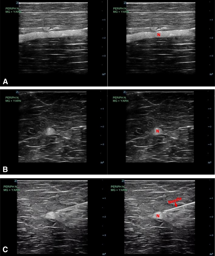
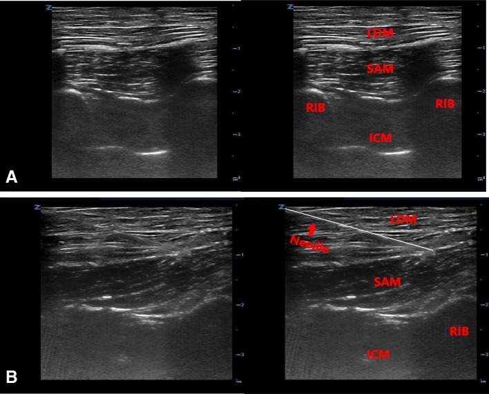
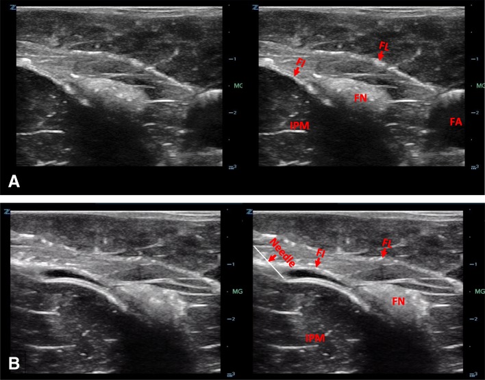
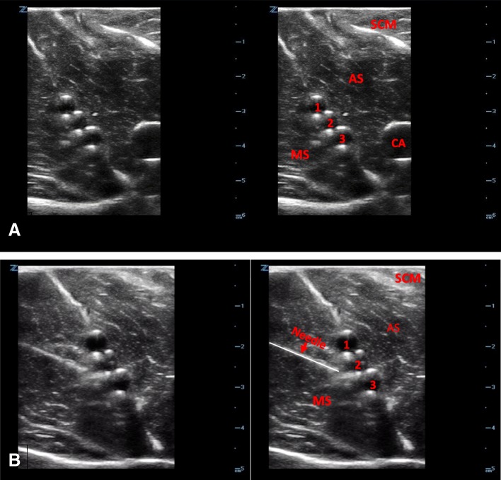
By using a lean cut of pork such as pork loin, yarn soaked in ultrasound gel to simulate peripheral nerves, and drinking straws filled with gel to represent vascular structures, we created various nerve block models. Meat glue applied between sections of meat appears hyperechoic under ultrasound, thereby mimicking fascial planes and has the added benefit of helping to secure the components of the model together. Using these elements, we were able to create realistic peripheral nerve, fascia iliaca compartment, serratus anterior plane, and interscalene brachial plexus models.
One of the necessary skills in performing UGRA involves placing the needle tip along a fascial plane and visualizing hydrodissection of this plane with the local anesthetic. When meat glue (transglutaminase) is applied between layers of meat such as pork loin, the meat binds together and creates a hyperechoic line that mimics a fascial plane. When meat glue is applied to two apposing fascial layers naturally occurring on the meat, the fascial plane can be injected, and fluid can be seen hydrodissecting in this space. We created several nerve block models using meat glue and other components to mimic normal landmarks.
We have developed inexpensive and easily reproducible models that create the realistic appearance of tissues, nerves, and fascial planes under ultrasound. They can also accurately simulate hydrodissection of fluid in fascial planes. We hope these nerve block models will allow for the education in UGRA to be more widespread and accessible to learners from all specialties.
超声引导区域麻醉(UGRA)越来越多地被急诊医师用于为患者提供安全有效的疼痛缓解。然而,限制其广泛应用的一个因素是缺乏可供学习者培训的现实模型。目前,尚无经济实惠的可注射神经阻滞模型,这些模型可模拟神经、筋膜平面、肌肉和其他解剖标志。我们的目标是创建经济实惠、可注射的神经阻滞模型,作为 UGRA 的有效医学培训工具。
我们使用像猪里脊肉这样的精瘦肉,将浸泡在超声凝胶中的纱线模拟外周神经,并用充满凝胶的吸管代表血管结构,从而创建了各种神经阻滞模型。肉胶涂在肉块之间,在超声下呈现高回声,从而模拟筋膜平面,并有助于将模型的各个部分固定在一起。使用这些元素,我们能够创建逼真的外周神经、髂筋膜间隙、前锯肌平面和锁骨下臂丛神经模型。
进行 UGRA 的必要技能之一是将针尖沿着筋膜平面放置,并使用局部麻醉剂可视化该平面的水分离。当肉胶(转谷氨酰胺酶)涂在猪里脊肉等肉块的层之间时,肉会粘在一起,形成一条高回声线,模拟筋膜平面。当肉胶涂在肉上自然存在的两个对向筋膜层上时,可以对筋膜平面进行注射,并可以看到在该空间内的流体水分离。我们使用肉胶和其他组件创建了几个神经阻滞模型,以模拟正常的解剖标志。
我们开发了经济实惠且易于复制的模型,这些模型在超声下创建了组织、神经和筋膜平面的逼真外观。它们还可以准确模拟流体在筋膜平面中的水分离。我们希望这些神经阻滞模型将使 UGRA 的教育更加广泛和易于为来自所有专业的学习者所接受。