Department of Radiology, the Affiliated Nanjing Drum Tower Hospital of Nanjing University Medical School, Nanjing 210008, China.
Research Center of Biostatistics and Computational Pharmacy, China Pharmaceutical University, Nanjing, China.
EBioMedicine. 2019 Jun;44:162-181. doi: 10.1016/j.ebiom.2019.05.040. Epub 2019 May 23.
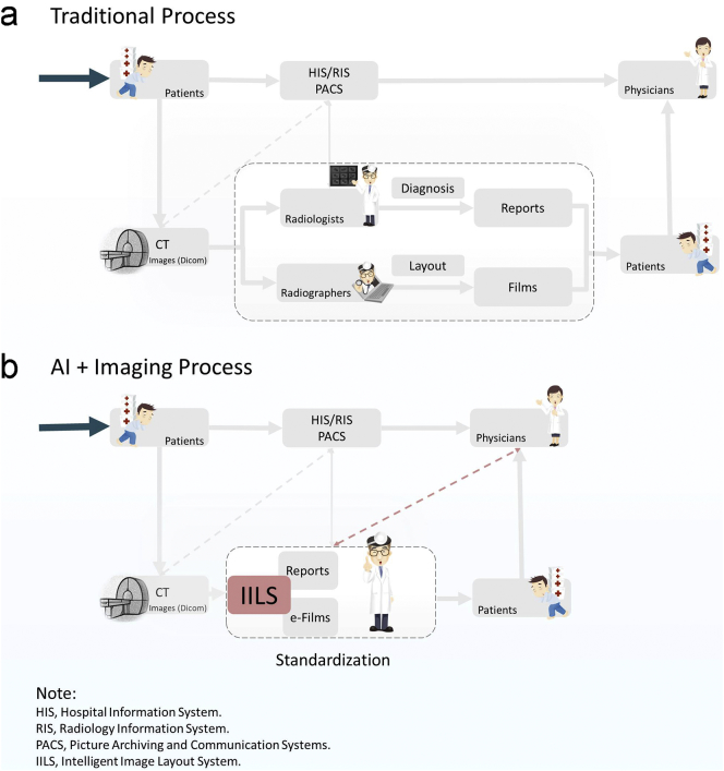
To achieve imaging report standardization and improve the quality and efficiency of the intra-interdisciplinary clinical workflow, we proposed an intelligent imaging layout system (IILS) for a clinical decision support system-based ubiquitous healthcare service, which is a lung nodule management system using medical images.
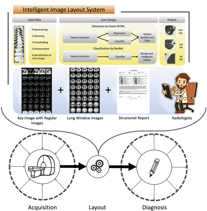
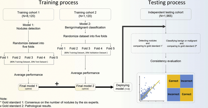
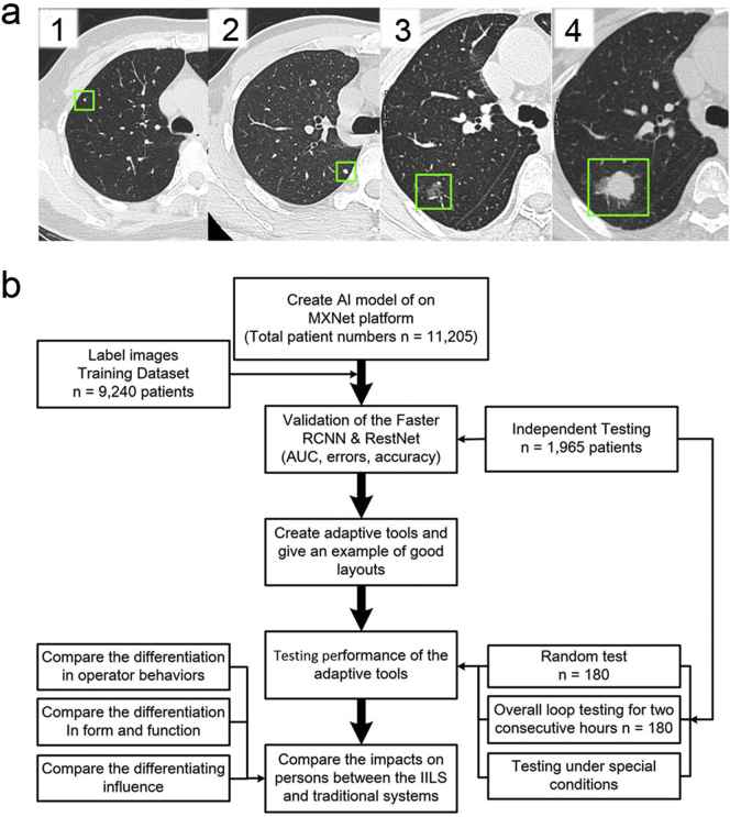
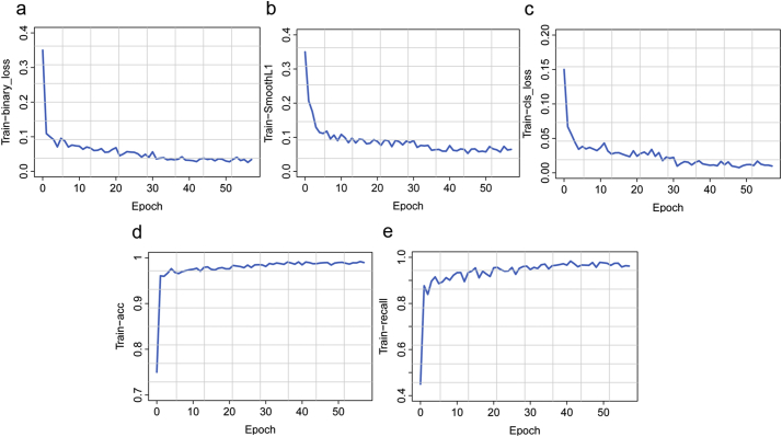
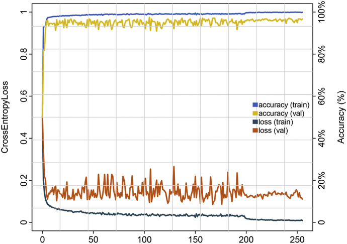
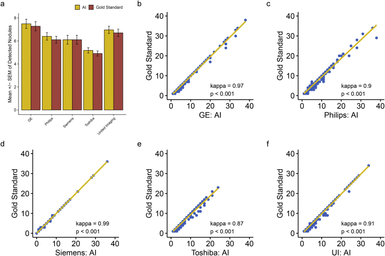
We created a lung IILS based on deep learning for imaging report standardization and workflow optimization for the identification of nodules. Our IILS utilized a deep learning plus adaptive auto layout tool, which trained and tested a neural network with imaging data from all the main CT manufacturers from 11,205 patients. Model performance was evaluated by the receiver operating characteristic curve (ROC) and calculating the corresponding area under the curve (AUC). The clinical application value for our IILS was assessed by a comprehensive comparison of multiple aspects.
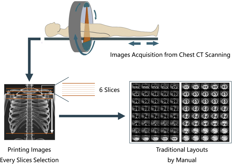
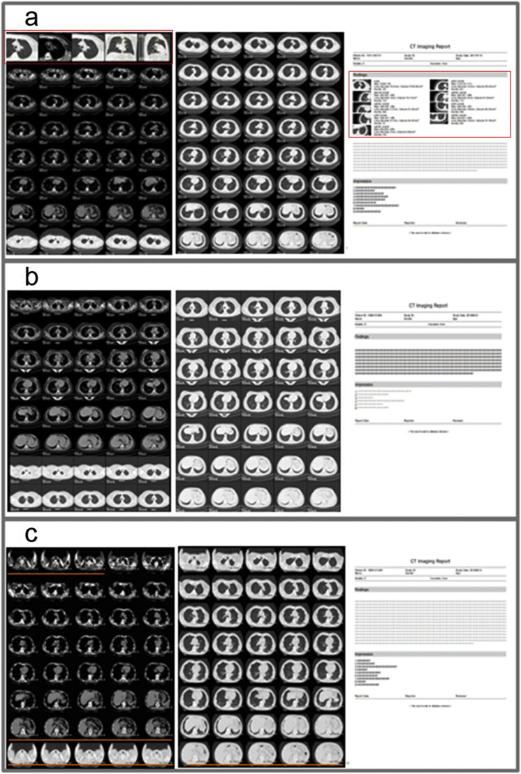
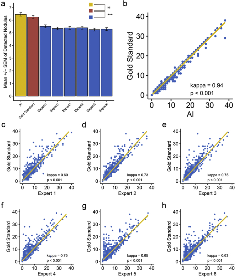
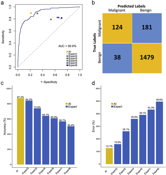
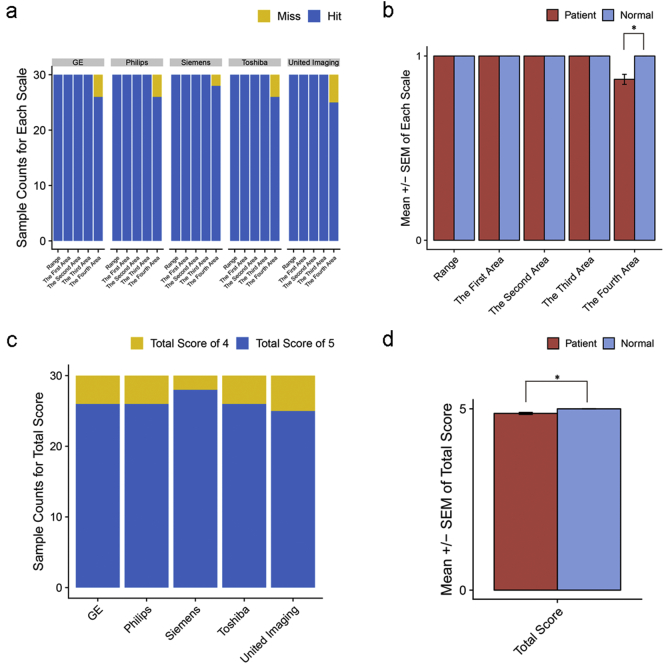
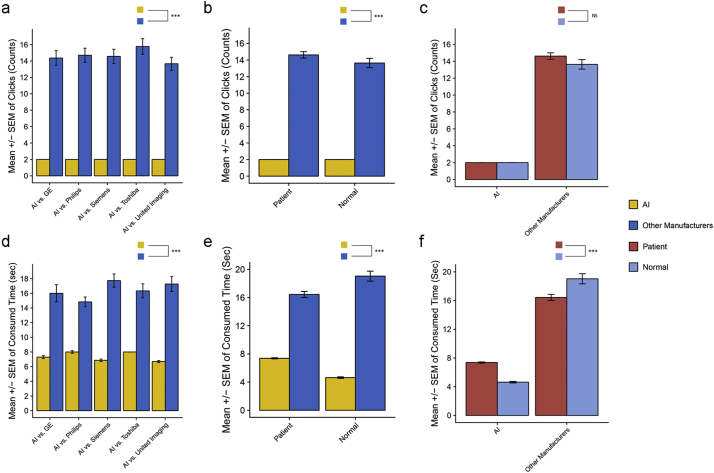
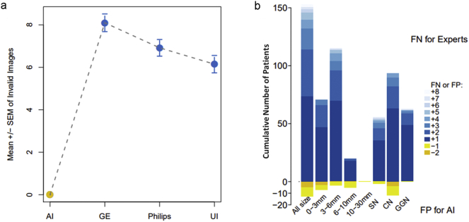
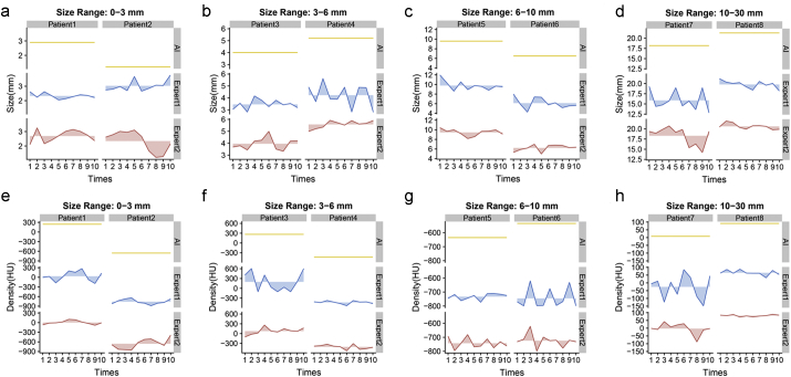
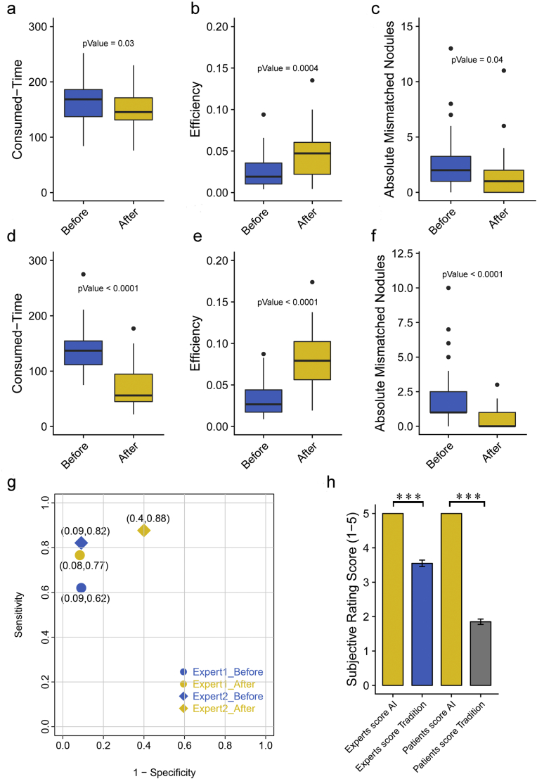
Our IILS is clinically applicable due to the consistency with nodules detected by IILS, with its highest consistency of 0·94 and an AUC of 90·6% for malignant pulmonary nodules versus benign nodules with a sensitivity of 76·5% and specificity of 89·1%. Applying this IILS to a dataset of chest CT images, we demonstrate performance comparable to that of human experts in providing a better layout and aiding in diagnosis in 100% valid images and nodule display. The IILS was superior to the traditional manual system in performance, such as reducing the number of clicks from 14·45 ± 0·38 to 2, time consumed from 16·87 ± 0·38 s to 6·92 ± 0·10 s, number of invalid images from 7·06 ± 0·24 to 0, and missing lung nodules from 46·8% to 0%.
This IILS might achieve imaging report standardization, and improve the clinical workflow therefore opening a new window for clinical application of artificial intelligence. FUND: The National Natural Science Foundation of China.
为了实现影像学报告标准化,提高跨学科临床工作流程的质量和效率,我们提出了一种基于临床决策支持系统的普遍医疗服务的智能影像学布局系统(IILS),这是一个用于管理肺部结节的医疗图像系统。
我们创建了一个基于深度学习的肺部 IILS,用于标准化影像学报告并优化识别结节的工作流程。我们的 IILS 利用了深度学习加自适应自动布局工具,该工具使用来自 11205 名患者的来自所有主要 CT 制造商的影像学数据对神经网络进行训练和测试。通过计算受试者工作特征曲线(ROC)及其相应的曲线下面积(AUC)来评估模型性能。通过对多个方面的综合比较,评估我们的 IILS 的临床应用价值。
由于 IILS 检测到的结节具有一致性,因此我们的 IILS 具有临床应用价值,其对恶性肺结节的最高一致性为 0.94,对良性结节的 AUC 为 90.6%,灵敏度为 76.5%,特异性为 89.1%。在对一组胸部 CT 图像数据集应用此 IILS 后,我们证明其在提供更好的布局和辅助诊断方面的性能可与人类专家媲美,并且在 100%有效图像和结节显示中都能够达到这一效果。与传统的手动系统相比,IILS 在性能方面具有优势,例如将点击次数从 14.45±0.38 减少到 2,将耗时从 16.87±0.38s 减少到 6.92±0.10s,将无效图像数量从 7.06±0.24 减少到 0,并将漏诊肺结节的比例从 46.8%降低到 0%。
该 IILS 可能实现影像学报告标准化,提高临床工作流程的效率,为人工智能在临床中的应用开辟新的窗口。
国家自然科学基金。