• 文献检索
  • 文档翻译
  • 深度研究
  • 学术资讯
  • Suppr Zotero 插件Zotero 插件
  • 邀请有礼
  • 套餐&价格
  • 历史记录
应用&插件
Suppr Zotero 插件Zotero 插件浏览器插件Mac 客户端Windows 客户端微信小程序
定价
高级版会员购买积分包购买API积分包
服务
文献检索文档翻译深度研究API 文档MCP 服务
关于我们
关于 Suppr公司介绍联系我们用户协议隐私条款
关注我们

Suppr 超能文献

核心技术专利:CN118964589B侵权必究
粤ICP备2023148730 号-1Suppr @ 2026

文献检索

告别复杂PubMed语法,用中文像聊天一样搜索,搜遍4000万医学文献。AI智能推荐,让科研检索更轻松。

立即免费搜索

文件翻译

保留排版,准确专业,支持PDF/Word/PPT等文件格式,支持 12+语言互译。

免费翻译文档

深度研究

AI帮你快速写综述,25分钟生成高质量综述,智能提取关键信息,辅助科研写作。

立即免费体验

乳腺光声成像:与组织病理学和组织病理生物标志物的相关性。

Optoacoustic imaging of the breast: correlation with histopathology and histopathologic biomarkers.

机构信息

Seno Medical Instruments, 8023 Vantage Dr. #1000, San Antonio, TX, 78230, USA.

Department of Radiology, Radboud University Medical Center, Geert Grooteplein Zuid 10, 6525 GA, Nijmegen, The Netherlands.

出版信息

Eur Radiol. 2019 Dec;29(12):6728-6740. doi: 10.1007/s00330-019-06262-0. Epub 2019 May 27.

DOI:10.1007/s00330-019-06262-0
PMID:31134367
原文链接:https://pmc.ncbi.nlm.nih.gov/articles/PMC6828639/
Abstract

AIM

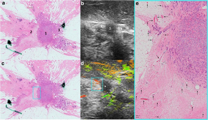
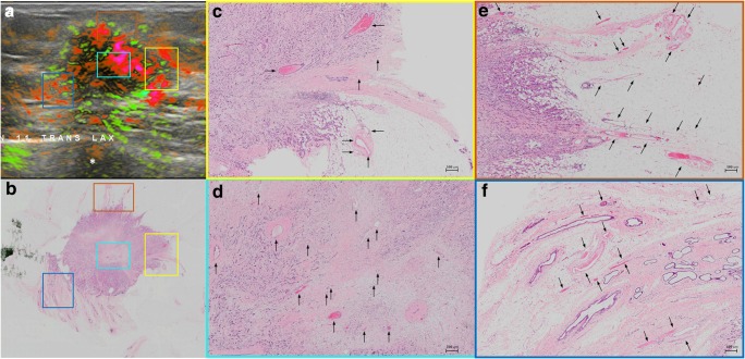
This study was conducted in order to investigate the role of gray-scale ultrasound (US) and optoacoustic imaging combined with gray-scale ultrasound (OA/US) to better differentiate between breast cancer molecular subtypes.

MATERIALS AND METHODS

All 67 malignant masses included in the Maestro trial were retrospectively reviewed to compare US and OA/US feature scores and histopathological findings. Kruskal-Wallis tests were used to analyze the relationship between US and OA/US features and molecular subtypes of breast cancer. If a significant relationship was found, additional Wilcoxon-Mann-Whitney tests were used to identify the differences between molecular subtype groups.

RESULTS

US sound transmission helped to differentiate between LUMA and LUMB, LUMB and TNBC, and LUMB and all other molecular subtypes combined (p values < 0.05). Regarding OA/US features, the sum of internal features helped to differentiate between TNBC and HER2-enriched subtypes (p = 0.049). Internal vessels (p = 0.025), sum of all internal features (p = 0.019), and sum of internal and external features (p = 0.028) helped to differentiate between LUMA and LUMB. All internal features, the sum of all internal features, the sum of all internal and external features, and the ratio of internal and external features helped to differentiate between LUMA and TNBC. The same features also helped to differentiate between LUMA and TNBC from other molecular subtypes (p values < 0.05).

CONCLUSIONS

The use of OA/US might help radiologists to better differentiate between breast cancer molecular subtypes. Further studies need to be carried out in order to validate these results.

KEY POINTS

• The combination of functional and morphologic information provided by optoacoustic imaging (OA) combined with gray-scale US helped to differentiate between breast cancer molecular subtypes.

摘要

目的

本研究旨在探讨灰阶超声(US)和光声成像(OA)联合灰阶超声(OA/US)在更好地区分乳腺癌分子亚型方面的作用。

材料与方法

回顾性分析 Maestro 试验中纳入的所有 67 个恶性肿块,比较 US 和 OA/US 特征评分与组织病理学结果。采用 Kruskal-Wallis 检验分析 US 和 OA/US 特征与乳腺癌分子亚型之间的关系。如果存在显著关系,则采用 Wilcoxon-Mann-Whitney 检验进一步比较各分子亚型组之间的差异。

结果

US 声传输有助于区分 LUMA 和 LUMB、LUMB 和 TNBC 以及 LUMB 和所有其他分子亚型的组合(p 值均<0.05)。OA/US 特征方面,内部特征之和有助于区分 TNBC 和 HER2 富集亚型(p=0.049)。内部血管(p=0.025)、所有内部特征之和(p=0.019)和内部及外部特征之和(p=0.028)有助于区分 LUMA 和 LUMB。所有内部特征、所有内部特征之和、所有内部和外部特征之和以及内部和外部特征之比有助于区分 LUMA 和 TNBC。同样的特征也有助于区分 LUMA 和 TNBC 与其他分子亚型(p 值均<0.05)。

结论

OA/US 的使用可能有助于放射科医生更好地区分乳腺癌分子亚型。需要进一步开展研究以验证这些结果。

关键要点

• 光声成像(OA)联合灰阶超声提供的功能和形态信息的组合有助于区分乳腺癌分子亚型。

https://cdn.ncbi.nlm.nih.gov/pmc/blobs/877b/6828639/c8876e64bd2b/330_2019_6262_Fig6_HTML.jpg
https://cdn.ncbi.nlm.nih.gov/pmc/blobs/877b/6828639/36a31aee02c3/330_2019_6262_Fig1_HTML.jpg
https://cdn.ncbi.nlm.nih.gov/pmc/blobs/877b/6828639/ff5603628e03/330_2019_6262_Fig2_HTML.jpg
https://cdn.ncbi.nlm.nih.gov/pmc/blobs/877b/6828639/5d4355d4837c/330_2019_6262_Fig3_HTML.jpg
https://cdn.ncbi.nlm.nih.gov/pmc/blobs/877b/6828639/416003277f65/330_2019_6262_Fig4_HTML.jpg
https://cdn.ncbi.nlm.nih.gov/pmc/blobs/877b/6828639/6f22b5df189d/330_2019_6262_Fig5_HTML.jpg
https://cdn.ncbi.nlm.nih.gov/pmc/blobs/877b/6828639/c8876e64bd2b/330_2019_6262_Fig6_HTML.jpg

相似文献

1
Optoacoustic imaging of the breast: correlation with histopathology and histopathologic biomarkers.乳腺光声成像:与组织病理学和组织病理生物标志物的相关性。
Eur Radiol. 2019 Dec;29(12):6728-6740. doi: 10.1007/s00330-019-06262-0. Epub 2019 May 27.
2
Optoacoustic Imaging and Gray-Scale US Features of Breast Cancers: Correlation with Molecular Subtypes.光声成像和灰阶超声在乳腺癌中的特征:与分子亚型的相关性。
Radiology. 2019 Sep;292(3):564-572. doi: 10.1148/radiol.2019182071. Epub 2019 Jul 9.
3
Optoacoustic Breast Imaging: Imaging-Pathology Correlation of Optoacoustic Features in Benign and Malignant Breast Masses.光声乳腺成像:良恶性乳腺肿块光声特征的成像-病理学相关性。
AJR Am J Roentgenol. 2018 Nov;211(5):1155-1170. doi: 10.2214/AJR.17.18435. Epub 2018 Aug 14.
4
Downgrading and Upgrading Gray-Scale Ultrasound BI-RADS Categories of Benign and Malignant Masses With Optoacoustics: A Pilot Study.超声 BI-RADS 灰阶良恶性肿块降级与升级与光声成像:一项初步研究。
AJR Am J Roentgenol. 2018 Sep;211(3):689-700. doi: 10.2214/AJR.17.18436. Epub 2018 Jul 5.
5
A Pivotal Study of Optoacoustic Imaging to Diagnose Benign and Malignant Breast Masses: A New Evaluation Tool for Radiologists.光声成像诊断良恶性乳腺肿块的关键研究:放射科医生的新评估工具。
Radiology. 2018 May;287(2):398-412. doi: 10.1148/radiol.2017172228. Epub 2017 Nov 27.
6
Downgrading of Breast Masses Suspicious for Cancer by Using Optoacoustic Breast Imaging.光声乳腺成像降低疑似乳腺癌的乳腺肿块的分级。
Radiology. 2018 Aug;288(2):355-365. doi: 10.1148/radiol.2018170500. Epub 2018 Apr 17.
7
Correlation between imaging features and molecular subtypes of breast cancer in young women (≤30 years old).年轻女性(≤30岁)乳腺癌的影像学特征与分子亚型之间的相关性
Jpn J Radiol. 2020 Nov;38(11):1062-1074. doi: 10.1007/s11604-020-01001-8. Epub 2020 Jun 19.
8
Contrast-enhanced cone beam breast CT features of breast cancers: correlation with immunohistochemical receptors and molecular subtypes.乳腺癌的对比增强锥形束乳腺CT特征:与免疫组化受体及分子亚型的相关性
Eur Radiol. 2021 Apr;31(4):2580-2589. doi: 10.1007/s00330-020-07277-8. Epub 2020 Oct 2.
9
Optoacoustic Imaging With Decision Support for Differentiation of Benign and Malignant Breast Masses: A 15-Reader Retrospective Study.基于决策支持的光声成像对良恶性乳腺肿块的鉴别诊断:一项 15 位读者的回顾性研究。
AJR Am J Roentgenol. 2023 May;220(5):646-658. doi: 10.2214/AJR.22.28470. Epub 2022 Dec 7.
10
Ultrasound-guided photoacoustic (US-PA) tomography of the breast: Biochemical differentiation using intrinsic tissue markers-lipids, collagen and hemoglobin with histopathologic correlation.超声引导光声(US-PA)断层扫描在乳腺中的应用:基于组织内在标志物——脂类、胶原和血红蛋白的生化差异与组织病理学相关性研究。
Sci Rep. 2024 Aug 5;14(1):18054. doi: 10.1038/s41598-024-65114-0.

引用本文的文献

1
Clinical and imaging characteristics of encapsulated papillary carcinoma: a comparative analysis with intraductal papilloma.包膜内乳头状癌的临床及影像学特征:与导管内乳头状瘤的对比分析
World J Surg Oncol. 2025 Jun 20;23(1):244. doi: 10.1186/s12957-025-03880-5.
2
Current status of optoacoustic breast imaging and future trends in clinical application: is it ready for prime time?光声乳腺成像的现状及临床应用的未来趋势:是否已准备好投入使用?
Eur Radiol. 2024 Sep;34(9):6092-6107. doi: 10.1007/s00330-024-10600-2. Epub 2024 Feb 3.
3
Optical Breast Imaging: A Review of Physical Principles, Technologies, and Clinical Applications.

本文引用的文献

1
ΔNp63 regulates the expression of hyaluronic acid-related genes in breast cancer cells.ΔNp63调节乳腺癌细胞中透明质酸相关基因的表达。
Oncogenesis. 2018 Aug 24;7(8):65. doi: 10.1038/s41389-018-0073-3.
2
Optoacoustic Breast Imaging: Imaging-Pathology Correlation of Optoacoustic Features in Benign and Malignant Breast Masses.光声乳腺成像:良恶性乳腺肿块光声特征的成像-病理学相关性。
AJR Am J Roentgenol. 2018 Nov;211(5):1155-1170. doi: 10.2214/AJR.17.18435. Epub 2018 Aug 14.
3
Downgrading of Breast Masses Suspicious for Cancer by Using Optoacoustic Breast Imaging.
光学乳腺成像:物理原理、技术及临床应用综述
J Breast Imaging. 2023 Sep-Oct;5(5):520-537. doi: 10.1093/jbi/wbad057. Epub 2023 Sep 22.
4
Photoacoustic imaging on its way toward clinical utility: a tutorial review focusing on practical application in medicine.光声成象迈向临床应用:聚焦于医学实际应用的教程综述
J Biomed Opt. 2023 Dec;28(12):121205. doi: 10.1117/1.JBO.28.12.121205. Epub 2023 Jun 8.
5
Understanding Breast Cancers through Spatial and High-Resolution Visualization Using Imaging Technologies.利用成像技术通过空间和高分辨率可视化来了解乳腺癌。
Cancers (Basel). 2022 Aug 23;14(17):4080. doi: 10.3390/cancers14174080.
6
Radiomics in Triple Negative Breast Cancer: New Horizons in an Aggressive Subtype of the Disease.三阴性乳腺癌中的放射组学:该侵袭性疾病亚型的新视野
J Clin Med. 2022 Jan 26;11(3):616. doi: 10.3390/jcm11030616.
7
Photoacoustic dual-scan mammoscope: results from 38 patients.光声双扫描乳腺镜:38例患者的结果
Biomed Opt Express. 2021 Mar 15;12(4):2054-2063. doi: 10.1364/BOE.420679. eCollection 2021 Apr 1.
8
Multi-modal imaging of high-risk ductal carcinoma in situ of the breast using C2Am: a targeted cell death imaging agent.使用 C2Am 对乳腺高危导管原位癌进行多模态成像:一种靶向细胞死亡成像剂。
Breast Cancer Res. 2021 Feb 17;23(1):25. doi: 10.1186/s13058-021-01404-z.
9
Integration of Multitargeted Polymer-Based Contrast Agents with Photoacoustic Computed Tomography: An Imaging Technique to Visualize Breast Cancer Intratumor Heterogeneity.多靶点聚合物基对比剂与光声计算机断层扫描的整合:一种可视化乳腺癌肿瘤内异质性的成像技术。
ACS Nano. 2021 Feb 23;15(2):2413-2427. doi: 10.1021/acsnano.0c05893. Epub 2021 Jan 19.
10
Opto-acoustic imaging of relative blood oxygen saturation and total hemoglobin for breast cancer diagnosis.用于乳腺癌诊断的相对血氧饱和度和总血红蛋白的光声成像。
J Biomed Opt. 2019 Dec;24(12):1-16. doi: 10.1117/1.JBO.24.12.121915.
光声乳腺成像降低疑似乳腺癌的乳腺肿块的分级。
Radiology. 2018 Aug;288(2):355-365. doi: 10.1148/radiol.2018170500. Epub 2018 Apr 17.
4
Breast Cancer Molecular Subtype Prediction by Mammographic Radiomic Features.基于乳腺影像组学特征的乳腺癌分子亚型预测。
Acad Radiol. 2019 Feb;26(2):196-201. doi: 10.1016/j.acra.2018.01.023. Epub 2018 Mar 8.
5
An inter-observer Ki67 reproducibility study applying two different assessment methods: on behalf of the Danish Scientific Committee of Pathology, Danish breast cancer cooperative group (DBCG).一项采用两种不同评估方法的观察者间Ki67重复性研究:代表丹麦病理学科学委员会、丹麦乳腺癌合作组(DBCG)开展。
Acta Oncol. 2018 Jan;57(1):83-89. doi: 10.1080/0284186X.2017.1404127. Epub 2017 Dec 5.
6
A Pivotal Study of Optoacoustic Imaging to Diagnose Benign and Malignant Breast Masses: A New Evaluation Tool for Radiologists.光声成像诊断良恶性乳腺肿块的关键研究:放射科医生的新评估工具。
Radiology. 2018 May;287(2):398-412. doi: 10.1148/radiol.2017172228. Epub 2017 Nov 27.
7
Predictive and prognostic impact of tumour-infiltrating lymphocytes in triple-negative breast cancer treated with neoadjuvant chemotherapy.新辅助化疗治疗三阴性乳腺癌中肿瘤浸润淋巴细胞的预测和预后影响
Ecancermedicalscience. 2017 Aug 15;11:759. doi: 10.3332/ecancer.2017.759. eCollection 2017.
8
MR Imaging Findings in Molecular Subtypes of Breast Cancer According to BIRADS System.根据乳腺影像报告和数据系统(BIRADS)的乳腺癌分子亚型的磁共振成像(MR)表现
Breast J. 2017 Jul;23(4):421-428. doi: 10.1111/tbj.12756. Epub 2017 Jan 9.
9
Pathological Characteristics of Triple-Negative Breast Cancer at Main Referral Teaching Hospital, April 2014 to April 2015, Tehran, Iran.2014年4月至2015年4月伊朗德黑兰主要转诊教学医院三阴性乳腺癌的病理特征
Int J Hematol Oncol Stem Cell Res. 2016 Oct 1;10(4):200-205.
10
Is There a Correlation between the Presence of a Spiculated Mass on Mammogram and Luminal A Subtype Breast Cancer?乳房X光片上出现的毛刺状肿块与腔面A型乳腺癌之间是否存在关联?
Korean J Radiol. 2016 Nov-Dec;17(6):846-852. doi: 10.3348/kjr.2016.17.6.846. Epub 2016 Oct 31.