• 文献检索
  • 文档翻译
  • 深度研究
  • 学术资讯
  • Suppr Zotero 插件Zotero 插件
  • 邀请有礼
  • 套餐&价格
  • 历史记录
应用&插件
Suppr Zotero 插件Zotero 插件浏览器插件Mac 客户端Windows 客户端微信小程序
定价
高级版会员购买积分包购买API积分包
服务
文献检索文档翻译深度研究API 文档MCP 服务
关于我们
关于 Suppr公司介绍联系我们用户协议隐私条款
关注我们

Suppr 超能文献

核心技术专利:CN118964589B侵权必究
粤ICP备2023148730 号-1Suppr @ 2026

文献检索

告别复杂PubMed语法,用中文像聊天一样搜索,搜遍4000万医学文献。AI智能推荐,让科研检索更轻松。

立即免费搜索

文件翻译

保留排版,准确专业,支持PDF/Word/PPT等文件格式,支持 12+语言互译。

免费翻译文档

深度研究

AI帮你快速写综述,25分钟生成高质量综述,智能提取关键信息,辅助科研写作。

立即免费体验

周边部视网膜内层变薄与路易体病的视觉功能障碍有关。

Parafoveal thinning of inner retina is associated with visual dysfunction in Lewy body diseases.

机构信息

Neurodegenerative Diseases Group, Biocruces Bizkaia Health Research Institute, Barakaldo, Spain.

Ophthalmology Department, Cruces University Hospital, Barakaldo, Spain.

出版信息

Mov Disord. 2019 Sep;34(9):1315-1324. doi: 10.1002/mds.27728. Epub 2019 May 28.

DOI:10.1002/mds.27728
PMID:31136022
原文链接:https://pmc.ncbi.nlm.nih.gov/articles/PMC6790692/
Abstract

BACKGROUND

Retinal optical coherence tomography findings in Lewy body diseases and their implications for visual outcomes remain controversial. We investigated whether region-specific thickness analysis of retinal layers could improve the detection of macular atrophy and unravel its association with visual disability in Parkinson's disease.

METHODS

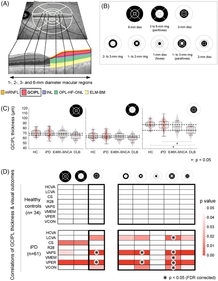
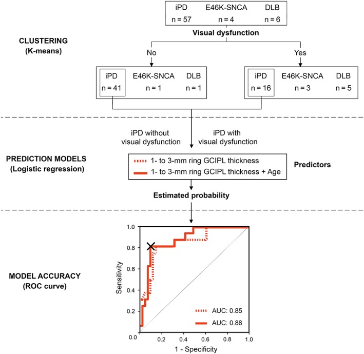
Patients with idiopathic Parkinson's disease (n = 63), dementia with Lewy bodies (n = 8), and E46K mutation carriers in the α-synuclein gene (E46K-SNCA) (n = 4) and 34 controls underwent Spectralis optical coherence tomography macular scans and a comprehensive battery of visual function and cognition tests. We computed mean retinal layer thicknesses of both eyes within 1-, 2-, 3-, and 6-mm diameter macular discs and in concentric parafoveal (1- to 2-mm, 2- to 3-mm, 1- to 3-mm) and perifoveal (3- to 6-mm) rings. Group differences in imaging parameters and their relationship with visual outcomes were analyzed. A multivariate logistic model was developed to predict visual impairment from optical coherence tomography measurements in Parkinson's disease, and cutoff values were determined with receiver operating characteristic analysis.

RESULTS

When compared with controls, patients with dementia with Lewy bodies had significant thinning of the ganglion cell-inner plexiform layer complex within the central 3-mm disc mainly because of differences in 1- to 3-mm parafoveal thickness. This parameter was strongly correlated in patients, but not in controls, with low contrast visual acuity and visual cognition outcomes (P < .05, False Discovery Rate), achieving 88% of accuracy in predicting visual impairment in Parkinson's disease.

CONCLUSION

Our findings support that parafoveal thinning of ganglion cell-inner plexiform complex is a sensitive and clinically relevant imaging biomarker for Lewy body diseases, specifically for Parkinson's disease. © 2019 The Authors. Movement Disorders published by Wiley Periodicals, Inc. on behalf of International Parkinson and Movement Disorder Society.

摘要

背景

Lewy 体疾病的视网膜光学相干断层扫描(OCT)结果及其对视功能预后的影响仍存在争议。我们旨在研究是否可以通过特定区域视网膜厚度分析来提高黄斑萎缩的检出率,并阐明其与帕金森病(PD)患者视力障碍的相关性。

方法

纳入特发性 PD 患者(63 例)、路易体痴呆(DLB)患者(8 例)、α-突触核蛋白基因 E46K 突变携带者(E46K-SNCA,4 例)和 34 名健康对照者,进行 Spectralis OCT 黄斑扫描和一系列视觉功能及认知测试。我们计算了双眼 1、2、3 和 6mm 黄斑盘直径内以及 1-2mm、2-3mm、1-3mm 近旁中心凹和 3-6mm 中心凹旁环的平均视网膜层厚度。分析了不同组别间的成像参数差异及其与视觉功能的相关性。建立了一个多变量逻辑回归模型,用于预测 PD 患者的 OCT 测量值与视力障碍的关系,并通过接受者操作特征曲线分析确定截断值。

结果

与对照组相比,DLB 患者的中央 3mm 盘内神经节细胞-内丛状层复合体厚度变薄,主要表现为 1-3mm 近旁中心凹厚度变薄。该参数在患者中与低对比视力和视觉认知结果密切相关(P<0.05,错误发现率),对 PD 患者的视力损害预测准确率达 88%。

结论

本研究结果支持神经节细胞-内丛状层复合体旁中心凹变薄是 Lewy 体疾病,尤其是 PD 的一种敏感且具有临床相关性的影像学生物标志物。

https://cdn.ncbi.nlm.nih.gov/pmc/blobs/09e9/6790692/4bee0752a3c6/MDS-34-1315-g003.jpg
https://cdn.ncbi.nlm.nih.gov/pmc/blobs/09e9/6790692/8a333d13a33a/MDS-34-1315-g001.jpg
https://cdn.ncbi.nlm.nih.gov/pmc/blobs/09e9/6790692/03929d946029/MDS-34-1315-g002.jpg
https://cdn.ncbi.nlm.nih.gov/pmc/blobs/09e9/6790692/4bee0752a3c6/MDS-34-1315-g003.jpg
https://cdn.ncbi.nlm.nih.gov/pmc/blobs/09e9/6790692/8a333d13a33a/MDS-34-1315-g001.jpg
https://cdn.ncbi.nlm.nih.gov/pmc/blobs/09e9/6790692/03929d946029/MDS-34-1315-g002.jpg
https://cdn.ncbi.nlm.nih.gov/pmc/blobs/09e9/6790692/4bee0752a3c6/MDS-34-1315-g003.jpg

相似文献

1
Parafoveal thinning of inner retina is associated with visual dysfunction in Lewy body diseases.周边部视网膜内层变薄与路易体病的视觉功能障碍有关。
Mov Disord. 2019 Sep;34(9):1315-1324. doi: 10.1002/mds.27728. Epub 2019 May 28.
2
Retinal Thickness Predicts the Risk of Cognitive Decline in Parkinson Disease.视网膜厚度可预测帕金森病患者认知能力下降的风险。
Ann Neurol. 2021 Jan;89(1):165-176. doi: 10.1002/ana.25944. Epub 2020 Nov 9.
3
Visual dysfunction and its correlation with retinal changes in patients with Parkinson's disease: an observational cross-sectional study.帕金森病患者的视觉功能障碍及其与视网膜变化的相关性:一项观察性横断面研究。
BMJ Open. 2016 May 6;6(5):e009658. doi: 10.1136/bmjopen-2015-009658.
4
Retina thickness as a marker of neurodegeneration in prodromal lewy body disease.视网膜厚度作为前驱路易体病神经退行性变的标志物。
Mov Disord. 2020 Feb;35(2):349-354. doi: 10.1002/mds.27914. Epub 2019 Nov 11.
5
Analysis of the retinal nerve fiber and ganglion cell - Inner plexiform layer by optical coherence tomography in Parkinson's patients.帕金森病患者视网膜神经纤维及神经节细胞-内网状层的光学相干断层扫描分析
Parkinsonism Relat Disord. 2016 Oct;31:59-64. doi: 10.1016/j.parkreldis.2016.07.004. Epub 2016 Jul 9.
6
Macular ganglion-cell-complex layer thinning and optic nerve integrity in drug-naïve Parkinson's disease.未经药物治疗的帕金森病的黄斑神经节细胞复合体层变薄与视神经完整性。
J Neural Transm (Vienna). 2019 Dec;126(12):1695-1699. doi: 10.1007/s00702-019-02097-7. Epub 2019 Oct 19.
7
Optical coherence tomography automated layer segmentation of macula after retinal detachment repair.视网膜脱离修复后黄斑的光学相干断层扫描自动分层。
PLoS One. 2018 May 7;13(5):e0197058. doi: 10.1371/journal.pone.0197058. eCollection 2018.
8
Diagnosis of Early-Stage Glaucoma by Grid-Wise Macular Inner Retinal Layer Thickness Measurement and Effect of Compensation of Disc-Fovea Inclination.通过网格状黄斑内层视网膜厚度测量诊断早期青光眼及视盘-黄斑倾斜补偿的效果
Invest Ophthalmol Vis Sci. 2015 Aug;56(9):5681-90. doi: 10.1167/iovs.15-17208.
9
Retinal nerve fiber layer thinning in dementia associated with Parkinson's disease, dementia with Lewy bodies, and Alzheimer's disease.帕金森病相关痴呆、路易体痴呆和阿尔茨海默病患者的视网膜神经纤维层变薄。
J Alzheimers Dis. 2013;34(3):659-64. doi: 10.3233/JAD-121975.
10
Alterations of the outer retina in non-arteritic anterior ischaemic optic neuropathy detected using spectral-domain optical coherence tomography.使用频域光学相干断层扫描检测非动脉炎性前部缺血性视神经病变中外层视网膜的改变。
Clin Exp Ophthalmol. 2017 Jul;45(5):496-508. doi: 10.1111/ceo.12914. Epub 2017 Mar 9.

引用本文的文献

1
Intrinsic capacity in Lewy body dementia: a review.路易体痴呆的内在能力:综述
J Frailty Aging. 2025 Aug;14(4):100076. doi: 10.1016/j.tjfa.2025.100076. Epub 2025 Aug 5.
2
Retinal Thickness in Patients with Parkinson's Disease and Dopa Responsive Dystonia-Is There Any Difference?帕金森病和多巴反应性肌张力障碍患者的视网膜厚度——有差异吗?
Biomedicines. 2025 May 19;13(5):1227. doi: 10.3390/biomedicines13051227.
3
Optical coherence tomography reveals retinal structural abnormalities in α-synucleinopathies: insights from the Padua-CESNE cohort.

本文引用的文献

1
Optical Coherence Tomography Findings in Patients of Parkinson's Disease: An Indian Perspective.帕金森病患者的光学相干断层扫描结果:印度视角
Ann Indian Acad Neurol. 2018 Apr-Jun;21(2):150-155. doi: 10.4103/aian.AIAN_152_18.
2
Retinal thinning associates with nigral dopaminergic loss in de novo Parkinson disease.视网膜变薄与新诊断帕金森病中的黑质多巴胺能神经元丧失有关。
Neurology. 2018 Sep 11;91(11):e1003-e1012. doi: 10.1212/WNL.0000000000006157. Epub 2018 Aug 15.
3
Retinal changes in Parkinson's disease and glaucoma.帕金森病和青光眼的视网膜变化。
光学相干断层扫描揭示α-突触核蛋白病中的视网膜结构异常:来自帕多瓦- CESNE队列的见解。
J Neural Transm (Vienna). 2025 Apr 15. doi: 10.1007/s00702-025-02918-y.
4
Amygdala Neurodegeneration: A Key Driver of Visual Dysfunction in Parkinson's Disease.杏仁核神经退行性变:帕金森病视觉功能障碍的关键驱动因素。
Ann Clin Transl Neurol. 2025 Apr;12(4):768-779. doi: 10.1002/acn3.70007. Epub 2025 Feb 17.
5
Dynamics of retinal changes in early-stage Parkinson's disease.早期帕金森病视网膜变化的动态研究
Acta Neuropathol Commun. 2025 Jan 31;13(1):20. doi: 10.1186/s40478-025-01936-x.
6
Anatomy-driven segmentation of parafoveal optical coherence tomography (OCT) measures may improve associations with clinical outcomes in multiple sclerosis.基于解剖学的黄斑旁光学相干断层扫描(OCT)测量分割可能会改善与多发性硬化症临床结局的相关性。
J Neurol. 2025 Jan 15;272(2):141. doi: 10.1007/s00415-024-12866-4.
7
pTau pathology in the retina of TAU58 mice: association with ganglion cell degeneration and implications on seeding and propagation of pTau from human brain lysates.TAU58小鼠视网膜中的pTau病理学:与神经节细胞变性的关联以及对来自人脑裂解物的pTau播种和传播的影响。
Acta Neuropathol Commun. 2024 Dec 20;12(1):194. doi: 10.1186/s40478-024-01907-8.
8
Neuroretinal and microvascular retinal features in dementia with Lewy body assessed by optical coherence tomography angiography.通过光学相干断层扫描血管造影评估路易体痴呆中的神经视网膜和视网膜微血管特征。
Neurol Sci. 2025 Jan;46(1):185-194. doi: 10.1007/s10072-024-07683-6. Epub 2024 Aug 17.
9
Macular structural integrity estimates are associated with Parkinson's disease genetic risk.黄斑结构完整性估计与帕金森病遗传风险相关。
Acta Neuropathol Commun. 2024 Aug 13;12(1):130. doi: 10.1186/s40478-024-01841-9.
10
Visual dysfunction in dementia with Lewy bodies.路易体痴呆的视觉功能障碍。
Curr Neurol Neurosci Rep. 2024 Aug;24(8):273-284. doi: 10.1007/s11910-024-01349-8. Epub 2024 Jun 22.
Parkinsonism Relat Disord. 2018 Nov;56:41-46. doi: 10.1016/j.parkreldis.2018.06.016. Epub 2018 Jun 21.
4
Phosphorylated α-synuclein in the retina is a biomarker of Parkinson's disease pathology severity.视网膜中磷酸化的α-突触核蛋白是帕金森病病理严重程度的生物标志物。
Mov Disord. 2018 Aug;33(8):1315-1324. doi: 10.1002/mds.27392. Epub 2018 May 8.
5
Progressive Changes in the Retinal Structure of Patients with Parkinson's Disease.帕金森病患者视网膜结构的进行性变化。
J Parkinsons Dis. 2018;8(1):85-92. doi: 10.3233/JPD-171184.
6
Correlations among multifocal electroretinography and optical coherence tomography findings in patients with Parkinson's disease.帕金森病患者多焦视网膜电图与光学相干断层扫描结果的相关性。
Neurol Sci. 2018 Mar;39(3):533-541. doi: 10.1007/s10072-018-3244-2. Epub 2018 Jan 18.
7
E46K α-synuclein pathological mutation causes cell-autonomous toxicity without altering protein turnover or aggregation.E46Kα-突触核蛋白病理性突变导致细胞自主性毒性,而不改变蛋白质周转或聚集。
Proc Natl Acad Sci U S A. 2017 Sep 26;114(39):E8274-E8283. doi: 10.1073/pnas.1703420114. Epub 2017 Sep 12.
8
Visual Dysfunction in Parkinson's Disease.帕金森病中的视觉功能障碍
Int Rev Neurobiol. 2017;134:921-946. doi: 10.1016/bs.irn.2017.04.007. Epub 2017 May 31.
9
Abnormal connectivity between the default mode and the visual system underlies the manifestation of visual hallucinations in Parkinson's disease: a task-based fMRI study.默认模式与视觉系统之间的异常连接是帕金森病视觉幻觉表现的基础:一项基于任务的功能磁共振成像研究。
NPJ Parkinsons Dis. 2015 Apr 22;1:15003. doi: 10.1038/npjparkd.2015.3. eCollection 2015.
10
Visual System Involvement in Patients with Newly Diagnosed Parkinson Disease.视觉系统在新发帕金森病患者中的受累情况。
Radiology. 2017 Dec;285(3):885-895. doi: 10.1148/radiol.2017161732. Epub 2017 Jul 11.