Kim Seung Soo, Kim Jae-Hun, Jeong Woo Kyoung, Lee Jisun, Kim Young Kon, Choi Dongil, Lee Won Jae
Department of Radiology and Center for Imaging Science, Samsung Medical Center, Sungkyunkwan University School of Medicine, Seoul.
Department of Radiology, Soonchunhyang University College of Medicine, Cheonan Hospital, Chungcheongnam-do.
Medicine (Baltimore). 2019 May;98(22):e15867. doi: 10.1097/MD.0000000000015867.
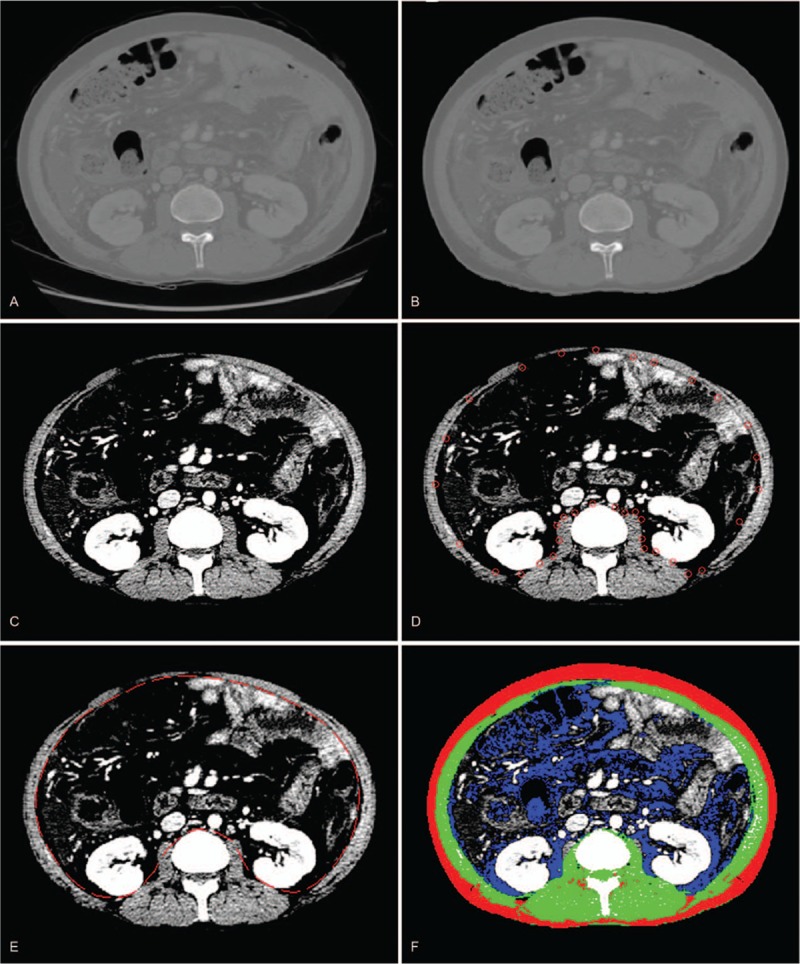
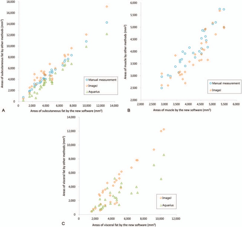
The aim of the study was to introduce our in-house software to measure the muscle and adipose area on axial computed tomography (CT) scans and to compare with various quantification methods.Our institutional review board approved this retrospective study and informed consent was waived. We developed in-house software to identify body composition analysis on CT scan, which semiautomatically operates 3 image processing steps. Abdominal images were obtained using multidetector row CT (MDCT). Two radiologists analyzed the same cross-sectional areas of subcutaneous fat, muscle, and visceral fat using the following techniques: manual measurements, Aquarius, ImageJ, and our newly developed software. We calculated an intraclass correlation coefficient (ICC) for comparison of muscle and fat areas quantified by various measurement methods using a 2-way random model. Interobserver agreement between the radiologists was also evaluated.Agreements in the measurement of subcutaneous fat and muscle areas were excellent among the methods (ICC = 0.962 and 0.897, respectively), and that of the visceral fat area was good (ICC = 0.822). In the subgroup analysis, ICC of the visceral fat area in the female group and in subjects with ascites was slightly lower than the other group (ICC = 0.742 and 0.787, respectively). The correlation coefficients between our software and other methods were relatively high (r = 0.854-0.996). Additionally, ICCs between both observers of our program for quantification of subcutaneous fat, muscle, and visceral fat areas were 0.999, 0.980, and 0.999, respectively.In conclusion, our method showed be reliable in quantifying muscle and adipose tissue using cross-sectional areas of MDCT with high reproducibility.
本研究的目的是介绍我们用于在轴向计算机断层扫描(CT)图像上测量肌肉和脂肪面积的内部软件,并与各种量化方法进行比较。我们的机构审查委员会批准了这项回顾性研究,且无需获得知情同意。我们开发了用于在CT扫描上进行身体成分分析的内部软件,该软件半自动地执行3个图像处理步骤。使用多排探测器CT(MDCT)获取腹部图像。两名放射科医生使用以下技术分析皮下脂肪、肌肉和内脏脂肪的相同横截面积:手动测量、Aquarius、ImageJ以及我们新开发的软件。我们使用双向随机模型计算组内相关系数(ICC),以比较通过各种测量方法量化的肌肉和脂肪面积。还评估了放射科医生之间的观察者间一致性。皮下脂肪和肌肉面积测量方法之间的一致性极佳(ICC分别为0.962和0.897),内脏脂肪面积测量方法之间的一致性良好(ICC = 0.822)。在亚组分析中,女性组和有腹水的受试者内脏脂肪面积的ICC略低于其他组(分别为ICC = 0.742和0.787)。我们的软件与其他方法之间的相关系数相对较高(r = 0.854 - 0.996)。此外,我们的程序中两名观察者对皮下脂肪、肌肉和内脏脂肪面积进行量化的ICC分别为0.999、0.980和0.999。总之,我们的方法在使用MDCT横截面积量化肌肉和脂肪组织方面显示出可靠性,且具有高重复性。