Conti Emilia, Allegra Mascaro Anna Letizia, Pavone Francesco Saverio
European Laboratory for Non-Linear Spectroscopy, University of Florence, 50019 Sesto Fiorentino, Italy.
Neuroscience Institute, National Research Council, 56100 Pisa, Italy.
Methods Protoc. 2019 Jan 29;2(1):11. doi: 10.3390/mps2010011.
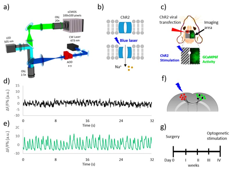
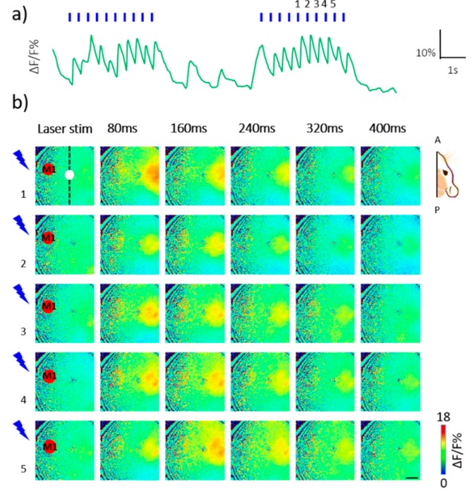
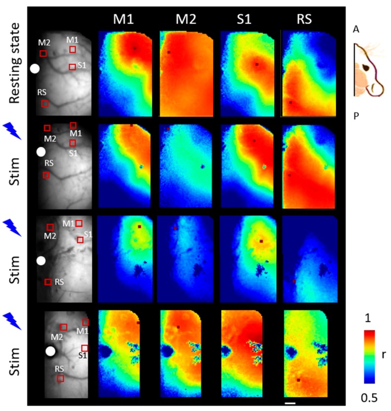
Recent improvements in optical tools that can perturb brain activity and simultaneously reveal the elicited alterations in the associated regions offer an exceptional means to understand and map the connectivity of the brain. In this work, we exploit a combination of recently developed optical tools to monitor neural population at the meso-scale level and to mould the cortical patterns of targeted neuronal population. Our goal was to investigate the propagation of neuronal activity over the mouse cortex that is triggered by optogenetic stimulation in the contralateral hemisphere. Towards this aim, we developed a wide-field fluorescence microscope that is characterized by a double illumination path allowing for the optogenetic stimulation of the transfected area in the left hemisphere and the simultaneous recording of cortical activity in the right hemisphere. The microscope was further implemented with a custom shutter in order to split the LED illumination path, resulting in a half-obscured field of view. By avoiding the spectral crosstalk between GCaMP6f and channelrhodopsin 2 (ChR2), this system offered the possibility of simultaneous "pumping and probing" of inter-hemispheric functional connectivity on Thy1-GCaMP6f mice.
近期,能够干扰大脑活动并同时揭示相关区域引发的变化的光学工具取得了进展,这为理解和绘制大脑的连接性提供了一种独特的方法。在这项工作中,我们利用最近开发的光学工具组合,在中尺度水平监测神经群体,并塑造目标神经元群体的皮质模式。我们的目标是研究由对侧半球的光遗传学刺激引发的神经元活动在小鼠皮质上的传播。为了实现这一目标,我们开发了一种宽场荧光显微镜,其特点是具有双照明路径,允许对左半球的转染区域进行光遗传学刺激,并同时记录右半球的皮质活动。该显微镜还配备了一个定制快门,以分割LED照明路径,从而产生一个半遮挡的视野。通过避免GCaMP6f和通道视紫红质2(ChR2)之间的光谱串扰,该系统提供了在Thy1-GCaMP6f小鼠上同时“激发和探测”半球间功能连接性的可能性。