• 文献检索
  • 文档翻译
  • 深度研究
  • 学术资讯
  • Suppr Zotero 插件Zotero 插件
  • 邀请有礼
  • 套餐&价格
  • 历史记录
应用&插件
Suppr Zotero 插件Zotero 插件浏览器插件Mac 客户端Windows 客户端微信小程序
定价
高级版会员购买积分包购买API积分包
服务
文献检索文档翻译深度研究API 文档MCP 服务
关于我们
关于 Suppr公司介绍联系我们用户协议隐私条款
关注我们

Suppr 超能文献

核心技术专利:CN118964589B侵权必究
粤ICP备2023148730 号-1Suppr @ 2026

文献检索

告别复杂PubMed语法,用中文像聊天一样搜索,搜遍4000万医学文献。AI智能推荐,让科研检索更轻松。

立即免费搜索

文件翻译

保留排版,准确专业,支持PDF/Word/PPT等文件格式,支持 12+语言互译。

免费翻译文档

深度研究

AI帮你快速写综述,25分钟生成高质量综述,智能提取关键信息,辅助科研写作。

立即免费体验

免疫胶体金 FIB-SEM:将体积超微结构可视化与 3D 生物分子分析相结合,以剖析细胞-环境相互作用。

Immunogold FIB-SEM: Combining Volumetric Ultrastructure Visualization with 3D Biomolecular Analysis to Dissect Cell-Environment Interactions.

机构信息

Department of Materials, Department of Bioengineering and Institute of Biomedical Engineering, Imperial College London, London, SW7 2AZ, UK.

Department of Medicine, Imperial College London, London, W12 0NN, UK.

出版信息

Adv Mater. 2019 Aug;31(32):e1900488. doi: 10.1002/adma.201900488. Epub 2019 Jun 13.

DOI:10.1002/adma.201900488
PMID:31197896
原文链接:https://pmc.ncbi.nlm.nih.gov/articles/PMC6778054/
Abstract

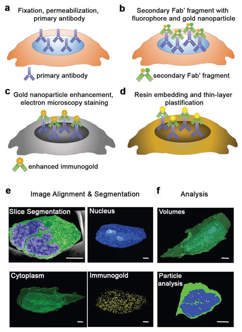
Volumetric imaging techniques capable of correlating structural and functional information with nanoscale resolution are necessary to broaden the insight into cellular processes within complex biological systems. The recent emergence of focused ion beam scanning electron microscopy (FIB-SEM) has provided unparalleled insight through the volumetric investigation of ultrastructure; however, it does not provide biomolecular information at equivalent resolution. Here, immunogold FIB-SEM, which combines antigen labeling with in situ FIB-SEM imaging, is developed in order to spatially map ultrastructural and biomolecular information simultaneously. This method is applied to investigate two different cell-material systems: the localization of histone epigenetic modifications in neural stem cells cultured on microstructured substrates and the distribution of nuclear pore complexes in myoblasts differentiated on a soft hydrogel surface. Immunogold FIB-SEM offers the potential for broad applicability to correlate structure and function with nanoscale resolution when addressing questions across cell biology, biomaterials, and regenerative medicine.

摘要

体积成像技术能够以纳米级分辨率关联结构和功能信息,对于深入了解复杂生物系统中的细胞过程至关重要。最近,聚焦离子束扫描电子显微镜(FIB-SEM)的出现通过对超微结构的体积研究提供了无与伦比的洞察力;然而,它无法以同等分辨率提供生物分子信息。在这里,开发了免疫金 FIB-SEM,它将抗原标记与原位 FIB-SEM 成像相结合,以便能够同时空间映射超微结构和生物分子信息。该方法应用于研究两个不同的细胞材料系统:在微结构基底上培养的神经干细胞中组蛋白表观遗传修饰的定位,以及在软水凝胶表面上分化的成肌细胞中核孔复合物的分布。免疫金 FIB-SEM 具有广泛的适用性,可在解决细胞生物学、生物材料和再生医学领域的问题时,以纳米级分辨率关联结构和功能。

https://cdn.ncbi.nlm.nih.gov/pmc/blobs/5003/6778054/b2871e830b67/EMS83593-f004.jpg
https://cdn.ncbi.nlm.nih.gov/pmc/blobs/5003/6778054/a9f36b39ad0a/EMS83593-f001.jpg
https://cdn.ncbi.nlm.nih.gov/pmc/blobs/5003/6778054/798ecaeed2e7/EMS83593-f002.jpg
https://cdn.ncbi.nlm.nih.gov/pmc/blobs/5003/6778054/3f6ce706172b/EMS83593-f003.jpg
https://cdn.ncbi.nlm.nih.gov/pmc/blobs/5003/6778054/b2871e830b67/EMS83593-f004.jpg

相似文献

1
Immunogold FIB-SEM: Combining Volumetric Ultrastructure Visualization with 3D Biomolecular Analysis to Dissect Cell-Environment Interactions.免疫胶体金 FIB-SEM:将体积超微结构可视化与 3D 生物分子分析相结合,以剖析细胞-环境相互作用。
Adv Mater. 2019 Aug;31(32):e1900488. doi: 10.1002/adma.201900488. Epub 2019 Jun 13.
2
Focused ion beam (FIB) combined with high resolution scanning electron microscopy: a promising tool for 3D analysis of chromosome architecture.聚焦离子束(FIB)与高分辨率扫描电子显微镜相结合:一种用于染色体结构三维分析的有前景的工具。
J Struct Biol. 2009 Feb;165(2):97-106. doi: 10.1016/j.jsb.2008.10.002. Epub 2008 Nov 5.
3
Volumetric 3D reconstruction of plant leaf cells using SEM, ion milling, TEM, and serial sectioning.使用 SEM、离子铣削、TEM 和连续切片对植物叶片细胞进行体积 3D 重建。
Planta. 2022 May 6;255(6):118. doi: 10.1007/s00425-022-03905-3.
4
Neuron Class and Target Variability in the Three-Dimensional Localization of SK2 Channels in Hippocampal Neurons as Detected by Immunogold FIB-SEM.免疫金标聚焦离子束扫描电子显微镜检测海马神经元中SK2通道三维定位的神经元类别和靶点变异性
Front Neuroanat. 2021 Dec 15;15:781314. doi: 10.3389/fnana.2021.781314. eCollection 2021.
5
FIB-SEM imaging properties of Drosophila melanogaster tissues embedded in Lowicryl HM20.在低聚甲醛 HM20 中包埋的果蝇组织的 FIB-SEM 成像特性。
J Microsc. 2019 Feb;273(2):91-104. doi: 10.1111/jmi.12764. Epub 2018 Nov 12.
6
Nanoscale Imaging of Collagen Gels with Focused Ion Beam Milling and Scanning Electron Microscopy.聚焦离子束铣削和扫描电子显微镜对胶原凝胶的纳米级成像
Biophys J. 2016 Oct 18;111(8):1797-1804. doi: 10.1016/j.bpj.2016.08.039.
7
Cryo-focused-ion-beam applications in structural biology.低温聚焦离子束在结构生物学中的应用。
Arch Biochem Biophys. 2015 Sep 1;581:122-30. doi: 10.1016/j.abb.2015.02.009. Epub 2015 Feb 20.
8
3D imaging of cells and tissues by focused ion beam/scanning electron microscopy (FIB/SEM).通过聚焦离子束/扫描电子显微镜(FIB/SEM)对细胞和组织进行三维成像。
Methods Mol Biol. 2013;950:275-92. doi: 10.1007/978-1-62703-137-0_16.
9
Combining serial block face and focused ion beam scanning electron microscopy for 3D studies of rare events.结合连续块面和聚焦离子束扫描电子显微镜用于罕见事件的三维研究。
Methods Cell Biol. 2019;152:87-101. doi: 10.1016/bs.mcb.2019.03.014. Epub 2019 May 7.
10
An optimized approach using cryofixation for high-resolution 3D analysis by FIB-SEM.利用 cryofixation 进行优化的方法,用于 FIB-SEM 的高分辨率 3D 分析。
J Struct Biol. 2020 Oct 1;212(1):107600. doi: 10.1016/j.jsb.2020.107600. Epub 2020 Aug 14.

引用本文的文献

1
Immunoelectron microscopy: a comprehensive guide from sample preparation to high-resolution imaging.免疫电子显微镜:从样品制备到高分辨率成像的全面指南
Discov Nano. 2025 Sep 8;20(1):154. doi: 10.1186/s11671-025-04346-z.
2
Microscopy methods for the in vivo study of nanoscale nuclear organization.用于纳米级细胞核组织体内研究的显微镜方法。
Biochem Soc Trans. 2025 Feb 3;53(1):BST20240629. doi: 10.1042/BST20240629.
3
silver nanoparticle development for molecular-specific biological imaging highly accessible microscopies.用于分子特异性生物成像的银纳米颗粒开发 高度可及的显微镜技术。

本文引用的文献

1
Accurate nanoelectrode recording of human pluripotent stem cell-derived cardiomyocytes for assaying drugs and modeling disease.用于药物检测和疾病建模的人类多能干细胞衍生心肌细胞的精确纳米电极记录。
Microsyst Nanoeng. 2017 Mar 13;3:16080. doi: 10.1038/micronano.2016.80. eCollection 2017.
2
Porous Silicon Nanoneedles Modulate Endocytosis to Deliver Biological Payloads.多孔硅纳米针调节内吞作用以递送生物有效载荷。
Adv Mater. 2019 Mar;31(12):e1806788. doi: 10.1002/adma.201806788. Epub 2019 Jan 24.
3
Engineering Anisotropic Muscle Tissue using Acoustic Cell Patterning.
Nanoscale Adv. 2022 Dec 21;5(6):1636-1650. doi: 10.1039/d2na00449f. eCollection 2023 Mar 14.
4
HP1 proteins regulate nucleolar structure and function by secluding pericentromeric constitutive heterochromatin.HP1 蛋白通过隔离着丝粒周围的组成型异染色质来调节核仁结构和功能。
Nucleic Acids Res. 2023 Jan 11;51(1):117-143. doi: 10.1093/nar/gkac1159.
5
Biophysical Regulations of Epigenetic State and Notch Signaling in Neural Development Using Microgroove Substrates.使用微槽基板对神经发育过程中表观遗传状态和Notch信号通路的生物物理调控
ACS Appl Mater Interfaces. 2022 Jul 13;14(29):32773-87. doi: 10.1021/acsami.2c01996.
6
Advancing cell instructive biomaterials through increased understanding of cell receptor spacing and material surface functionalization.通过加深对细胞受体间距和材料表面功能化的理解来推进细胞诱导生物材料的发展。
Regen Eng Transl Med. 2021 Dec;7(4):553-547. doi: 10.1007/s40883-020-00180-0. Epub 2020 Nov 20.
7
Electron Microscopy Techniques for Investigating Structure and Composition of Hair-Cell Stereociliary Bundles.用于研究毛细胞静纤毛束结构与组成的电子显微镜技术
Front Cell Dev Biol. 2021 Oct 22;9:744248. doi: 10.3389/fcell.2021.744248. eCollection 2021.
8
Direct imaging of antigen-antibody binding by atomic force microscopy.通过原子力显微镜对抗原-抗体结合进行直接成像。
Appl Nanosci. 2021;11(1):293-300. doi: 10.1007/s13204-020-01558-w. Epub 2020 Sep 24.
9
Volume electron microscopy: analyzing the lung.体视电子显微镜术:分析肺部
Histochem Cell Biol. 2021 Feb;155(2):241-260. doi: 10.1007/s00418-020-01916-3. Epub 2020 Sep 17.
10
High-Aspect-Ratio Nanostructured Surfaces as Biological Metamaterials.高纵横比纳米结构表面:生物类质材料
Adv Mater. 2020 Mar;32(9):e1903862. doi: 10.1002/adma.201903862. Epub 2020 Jan 16.
利用声控细胞图案化技术构建各向异性肌肉组织
Adv Mater. 2018 Oct;30(43):e1802649. doi: 10.1002/adma.201802649. Epub 2018 Sep 12.
4
Nanoscale invaginations of the nuclear envelope: Shedding new light on wormholes with elusive function.核膜的纳米级内陷:为具有难以捉摸功能的虫孔带来新的曙光。
Nucleus. 2017 Sep 3;8(5):506-514. doi: 10.1080/19491034.2017.1337621. Epub 2017 Jul 7.
5
Revealing the Cell-Material Interface with Nanometer Resolution by Focused Ion Beam/Scanning Electron Microscopy.利用聚焦离子束/扫描电子显微镜以纳米分辨率揭示细胞-材料界面。
ACS Nano. 2017 Aug 22;11(8):8320-8328. doi: 10.1021/acsnano.7b03494. Epub 2017 Jul 21.
6
Direct Evidence of Lack of Colocalisation of Fluorescently Labelled Gold Labels Used in Correlative Light Electron Microscopy.直接证据表明,在相关光电子显微镜中使用的荧光标记金标记物不存在共定位。
Sci Rep. 2017 Mar 20;7:44666. doi: 10.1038/srep44666.
7
Compression Induced Chondrogenic Differentiation of Embryonic Stem Cells in Three-Dimensional Polydimethylsiloxane Scaffolds.三维聚二甲基硅氧烷支架中压缩诱导胚胎干细胞的软骨分化
Tissue Eng Part A. 2017 May;23(9-10):426-435. doi: 10.1089/ten.TEA.2016.0376. Epub 2017 Feb 24.
8
Imaging cellular structures in super-resolution with SIM, STED and Localisation Microscopy: A practical comparison.利用结构照明显微镜(SIM)、受激发射损耗显微镜(STED)和定位显微镜进行超分辨率细胞结构成像:实际比较
Sci Rep. 2016 Jun 6;6:27290. doi: 10.1038/srep27290.
9
H3K9me3-Dependent Heterochromatin: Barrier to Cell Fate Changes.H3K9me3依赖的异染色质:细胞命运改变的障碍。
Trends Genet. 2016 Jan;32(1):29-41. doi: 10.1016/j.tig.2015.11.001. Epub 2015 Dec 8.
10
Chromatin states and nuclear organization in development--a view from the nuclear lamina.发育过程中的染色质状态与核组织——从核纤层角度的观察
Genome Biol. 2015 Aug 25;16(1):174. doi: 10.1186/s13059-015-0747-5.