From the Centre for Prevention of Stroke and Dementia (G.Z., L.G., S.M., S.T.P., P.M.R.) and Wellcome Centre for Integrative Neuroimaging, FMRIB (G.Z., L.G.), Nuffield Department of Clinical Neurosciences, John Radcliffe Hospital, University of Oxford; and Department of Biomedical, Metabolic and Neural Sciences and Centre for Neurosciences and Neurotechnology (G.Z.), University of Modena and Reggio Emilia, Italy.
Neurology. 2019 Jul 16;93(3):e272-e282. doi: 10.1212/WNL.0000000000007772. Epub 2019 Jun 14.
To investigate if the association between MRI-detectable white matter hyperintensity (WMH) and cognitive status reported in previous studies persists at older ages (>80 years), when some white matter abnormality is almost universally reported in clinical practice.
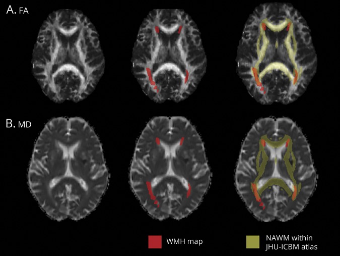
Consecutive eligible patients from a population-based cohort of all TIA/nondisabling stroke (Oxford Vascular Study) underwent multimodal MRI, including fluid-attenuated inversion recovery and diffusion-weighted imaging, allowing automated measurement of WMH volume, mean diffusivity (MD), and fractional anisotropy (FA) in normal-appearing white matter using FSL tools. These measures were related to cognitive status (Montreal Cognitive Assessment) at age ≤80 vs >80 years.
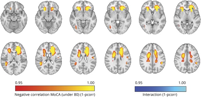
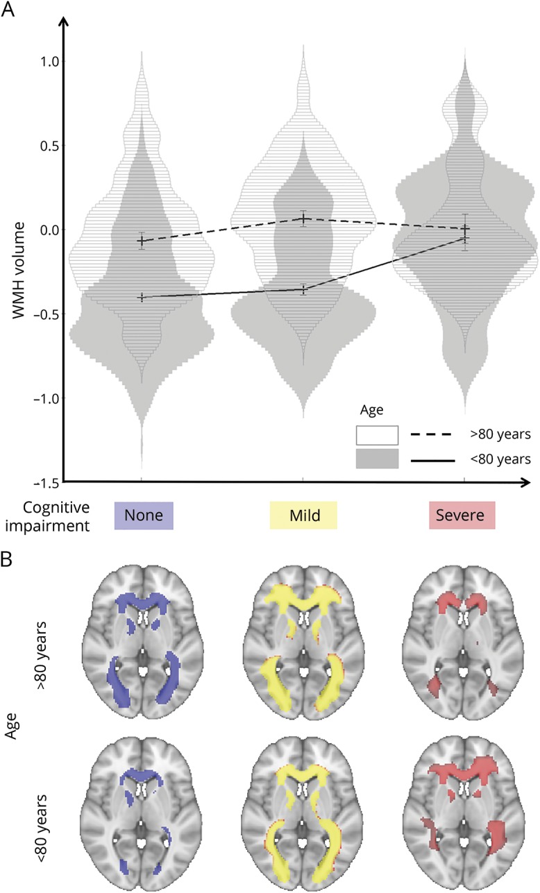
Of 566 patients (mean [range] age 66.7 [20-102] years), 107 were aged >80 years. WMH volumes and MD/FA were strongly associated with cognitive status in patients aged ≤80 years (all < 0.001 for WMH, MD, and FA) but not in patients aged >80 years (not significant for WMH, MD, and FA), with age interactions for WMH volume ( = 0.016) and MD ( = 0.037). Voxel-wise analyses also showed that lower Montreal Cognitive Assessment scores were associated with frontal WMH in patients ≤80 years, but not >80 years.
MRI markers of white matter damage are strongly related to cognition in patients with TIA/minor stroke at younger ages, but not at age >80 years. Clinicians and patients should not overinterpret the significance of these abnormalities at older ages.
探究在高龄(>80 岁)人群中,当一些脑白质异常在临床实践中几乎普遍存在时,先前研究报告的 MRI 可检测到的脑白质高信号(WMH)与认知状态之间的关联是否仍然存在。
来自一项基于人群的短暂性脑缺血发作/非致残性中风队列(牛津血管研究)的连续合格患者接受了多模态 MRI 检查,包括液体衰减反转恢复和弥散加权成像,使用 FSL 工具可以自动测量正常表现白质中的 WMH 体积、平均弥散度(MD)和各向异性分数(FA)。这些指标与≤80 岁和>80 岁时的认知状态(蒙特利尔认知评估)相关。
在 566 名患者(平均[范围]年龄 66.7[20-102]岁)中,有 107 名患者年龄>80 岁。WMH 体积和 MD/FA 与≤80 岁患者的认知状态密切相关(WMH、MD 和 FA 的所有指标均<0.001),但与>80 岁患者无关(WMH、MD 和 FA 均无统计学意义),WMH 体积( = 0.016)和 MD( = 0.037)存在年龄交互作用。体素分析还显示,在≤80 岁的患者中,较低的蒙特利尔认知评估评分与额部 WMH 相关,但在>80 岁的患者中则没有。
在年轻患者(TIA/轻度中风)中,MRI 脑白质损伤标志物与认知功能密切相关,但在>80 岁的患者中则没有。临床医生和患者不应在高龄时过度解读这些异常的意义。