• 文献检索
  • 文档翻译
  • 深度研究
  • 学术资讯
  • Suppr Zotero 插件Zotero 插件
  • 邀请有礼
  • 套餐&价格
  • 历史记录
应用&插件
Suppr Zotero 插件Zotero 插件浏览器插件Mac 客户端Windows 客户端微信小程序
定价
高级版会员购买积分包购买API积分包
服务
文献检索文档翻译深度研究API 文档MCP 服务
关于我们
关于 Suppr公司介绍联系我们用户协议隐私条款
关注我们

Suppr 超能文献

核心技术专利:CN118964589B侵权必究
粤ICP备2023148730 号-1Suppr @ 2026

文献检索

告别复杂PubMed语法,用中文像聊天一样搜索,搜遍4000万医学文献。AI智能推荐,让科研检索更轻松。

立即免费搜索

文件翻译

保留排版,准确专业,支持PDF/Word/PPT等文件格式,支持 12+语言互译。

免费翻译文档

深度研究

AI帮你快速写综述,25分钟生成高质量综述,智能提取关键信息,辅助科研写作。

立即免费体验

基于人工智能的脑肿瘤周围微环境局部标志物。

Artificial intelligence-based locoregional markers of brain peritumoral microenvironment.

机构信息

Diffusion & Connectomics In Precision Healthcare Research (DiCIPHR) Lab, University of Pennsylvania, Philadelphia, PA, 19104, USA.

Center for Biomedical Image Computing and Analytics (CBICA), University of Pennsylvania, Philadelphia, PA, 19104, USA.

出版信息

Sci Rep. 2023 Jan 18;13(1):963. doi: 10.1038/s41598-022-26448-9.

DOI:10.1038/s41598-022-26448-9
PMID:36653382
原文链接:https://pmc.ncbi.nlm.nih.gov/articles/PMC9849348/
Abstract

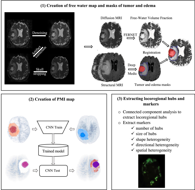
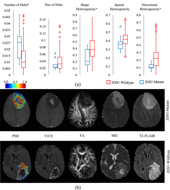
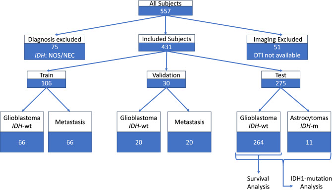
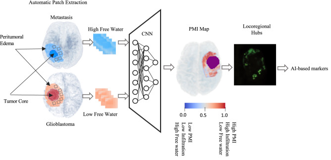
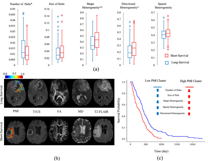
In malignant primary brain tumors, cancer cells infiltrate into the peritumoral brain structures which results in inevitable recurrence. Quantitative assessment of infiltrative heterogeneity in the peritumoral region, the area where biopsy or resection can be hazardous, is important for clinical decision making. Here, we derive a novel set of Artificial intelligence (AI)-based markers capturing the heterogeneity of tumor infiltration, by characterizing free water movement restriction in the peritumoral region using Diffusion Tensor Imaging (DTI)-based free water volume fraction maps. We leverage the differences in the peritumoral region of metastasis and glioblastomas, the former consisting of vasogenic versus the latter containing infiltrative edema, to extract a voxel-wise deep learning-based peritumoral microenvironment index (PMI). Descriptive characteristics of locoregional hubs of uniformly high PMI values are then extracted as AI-based markers to capture distinct aspects of infiltrative heterogeneity. The proposed markers are utilized to stratify patients' survival and IDH1 mutation status on a population of 275 adult-type diffuse gliomas (CNS WHO grade 4). Our results show significant differences in the proposed markers between patients with different overall survival and IDH1 mutation status (t test, Wilcoxon rank sum test, linear regression; p < 0.01). Clustering of patients using the proposed markers reveals distinct survival groups (logrank; p < 10, Cox hazard ratio = 1.82; p < 0.005). Our findings provide a panel of markers as surrogates of infiltration that might capture novel insight about underlying biology of peritumoral microstructural heterogeneity, providing potential biomarkers of prognosis pertaining to survival and molecular stratification, with applicability in clinical decision making.

摘要

在恶性原发性脑肿瘤中,癌细胞浸润到肿瘤周围的脑结构中,导致不可避免的复发。对肿瘤周围区域浸润异质性的定量评估,即活检或切除可能有危险的区域,对临床决策至关重要。在这里,我们通过使用基于弥散张量成像(DTI)的自由水体积分数图来描述肿瘤周围区域的自由水运动受限,从而得出一组新的基于人工智能(AI)的标志物,这些标志物可以捕捉肿瘤浸润的异质性。我们利用转移瘤和胶质母细胞瘤(前者由血管源性组成,后者包含浸润性水肿)在肿瘤周围区域的差异,提取基于体素的深度学习肿瘤周围微环境指数(PMI)。然后,提取局部区域高均一 PMI 值的神经中枢的描述性特征作为 AI 标志物,以捕获浸润异质性的不同方面。所提出的标志物用于对 275 名成人型弥漫性神经胶质瘤(CNS WHO 分级 4)患者的生存和 IDH1 突变状态进行分层。我们的研究结果表明,在不同总生存和 IDH1 突变状态的患者中,所提出的标志物存在显著差异(t 检验、Wilcoxon 秩和检验、线性回归;p<0.01)。使用所提出的标志物对患者进行聚类,揭示了不同的生存组(logrank;p<10,Cox 风险比=1.82;p<0.005)。我们的研究结果提供了一组作为浸润替代物的标志物,这些标志物可能提供有关肿瘤周围微结构异质性潜在生物学的新见解,提供与生存和分子分层相关的预后潜在生物标志物,适用于临床决策。

https://cdn.ncbi.nlm.nih.gov/pmc/blobs/1157/9849348/b484f6c19e35/41598_2022_26448_Fig6_HTML.jpg
https://cdn.ncbi.nlm.nih.gov/pmc/blobs/1157/9849348/2518f2746bf1/41598_2022_26448_Fig1_HTML.jpg
https://cdn.ncbi.nlm.nih.gov/pmc/blobs/1157/9849348/aab36dfa11ff/41598_2022_26448_Fig2_HTML.jpg
https://cdn.ncbi.nlm.nih.gov/pmc/blobs/1157/9849348/95e4be65ff32/41598_2022_26448_Fig3_HTML.jpg
https://cdn.ncbi.nlm.nih.gov/pmc/blobs/1157/9849348/3731dcf1aa42/41598_2022_26448_Fig4_HTML.jpg
https://cdn.ncbi.nlm.nih.gov/pmc/blobs/1157/9849348/074d7ac5bd31/41598_2022_26448_Fig5_HTML.jpg
https://cdn.ncbi.nlm.nih.gov/pmc/blobs/1157/9849348/b484f6c19e35/41598_2022_26448_Fig6_HTML.jpg

相似文献

1
Artificial intelligence-based locoregional markers of brain peritumoral microenvironment.基于人工智能的脑肿瘤周围微环境局部标志物。
Sci Rep. 2023 Jan 18;13(1):963. doi: 10.1038/s41598-022-26448-9.
2
Distinct tumor signatures using deep learning-based characterization of the peritumoral microenvironment in glioblastomas and brain metastases.基于深度学习的脑胶质瘤和脑转移瘤瘤周微环境特征分析,揭示肿瘤异质性特征。
Sci Rep. 2021 Jul 14;11(1):14469. doi: 10.1038/s41598-021-93804-6.
3
Advanced diffusion MRI provides evidence for altered axonal microstructure and gradual peritumoral infiltration in GBM in comparison to brain metastases.高级弥散 MRI 为胶质母细胞瘤与脑转移瘤相比,存在轴突微观结构改变和逐渐发生的肿瘤周围浸润提供了证据。
Clin Neuroradiol. 2024 Sep;34(3):703-711. doi: 10.1007/s00062-024-01416-0. Epub 2024 Apr 29.
4
The differences of water diffusion between brain tissue infiltrated by tumor and peritumoral vasogenic edema.肿瘤浸润脑组织与瘤周血管源性水肿之间水扩散的差异。
Clin Imaging. 2009 Mar-Apr;33(2):96-101. doi: 10.1016/j.clinimag.2008.06.035.
5
Quantitative apparent diffusion coefficients in the characterization of brain tumors and associated peritumoral edema.定量表观扩散系数在脑肿瘤及相关瘤周水肿特征描述中的应用
Acta Radiol. 2009 Jul;50(6):682-9. doi: 10.1080/02841850902933123.
6
Texture analysis on diffusion tensor imaging: discriminating glioblastoma from single brain metastasis.扩散张量成像的纹理分析:鉴别胶质母细胞瘤与单发脑转移瘤。
Acta Radiol. 2019 Mar;60(3):356-366. doi: 10.1177/0284185118780889. Epub 2018 Jun 3.
7
Diffusion tensor imaging provides an insight into the microstructure of meningiomas, high-grade gliomas, and peritumoral edema.扩散张量成像有助于深入了解脑膜瘤、高级别胶质瘤和瘤周水肿的微观结构。
J Comput Assist Tomogr. 2012 Sep-Oct;36(5):577-82. doi: 10.1097/RCT.0b013e318261e913.
8
Peritumoral diffusion tensor imaging of high-grade gliomas and metastatic brain tumors.高级别胶质瘤和脑转移瘤的瘤周扩散张量成像
AJNR Am J Neuroradiol. 2003 May;24(5):937-41.
9
Differentiation of brain tumor-related edema based on 3D T1rho imaging.基于三维T1rho成像的脑肿瘤相关水肿的鉴别
Eur J Radiol. 2017 Jun;91:88-92. doi: 10.1016/j.ejrad.2017.03.022. Epub 2017 Mar 30.
10
Imaging ¹⁸F-fluorodeoxy glucose/¹¹C-methionine uptake decoupling for identification of tumor cell infiltration in peritumoral brain edema.¹⁸F-氟脱氧葡萄糖/¹¹C-蛋氨酸摄取解偶联成像用于识别瘤周脑水肿中的肿瘤细胞浸润。
J Neurooncol. 2012 Jan;106(2):417-25. doi: 10.1007/s11060-011-0688-0. Epub 2011 Aug 17.

引用本文的文献

1
Diagnostic, Therapeutic, and Prognostic Applications of Artificial Intelligence (AI) in the Clinical Management of Brain Metastases (BMs).人工智能(AI)在脑转移瘤(BMs)临床管理中的诊断、治疗及预后应用
Brain Sci. 2025 Jul 8;15(7):730. doi: 10.3390/brainsci15070730.
2
Individual Brain Tumor Invasion Mapping Based on Diffusion Kurtosis Imaging.基于扩散峰度成像的个体脑肿瘤侵袭图谱
Sovrem Tekhnologii Med. 2025;17(1):81-90. doi: 10.17691/stm2025.17.1.08. Epub 2025 Feb 28.
3
Exploring the roles and clinical potential of exosome-derived non-coding RNAs in glioma.

本文引用的文献

1
Glioma invasion along white matter tracts: A dilemma for neurosurgeons.胶质瘤沿白质纤维束侵袭:神经外科医生面临的困境。
Cancer Lett. 2022 Feb 1;526:103-111. doi: 10.1016/j.canlet.2021.11.020. Epub 2021 Nov 20.
2
Distinct tumor signatures using deep learning-based characterization of the peritumoral microenvironment in glioblastomas and brain metastases.基于深度学习的脑胶质瘤和脑转移瘤瘤周微环境特征分析,揭示肿瘤异质性特征。
Sci Rep. 2021 Jul 14;11(1):14469. doi: 10.1038/s41598-021-93804-6.
3
Hypermetabolism and impaired cerebrovascular reactivity beyond the standard MRI-identified tumor border indicate diffuse glioma extended tissue infiltration.
探索外泌体来源的非编码RNA在胶质瘤中的作用及临床潜力。
IBRO Neurosci Rep. 2025 Feb 5;18:323-337. doi: 10.1016/j.ibneur.2025.01.015. eCollection 2025 Jun.
4
Vagus nerve stimulation: Novel concept for the treatment of glioblastoma and solid cancers by cytokine (interleukin-6) reduction, attenuating the SASP, enhancing tumor immunity.迷走神经刺激:通过降低细胞因子(白细胞介素-6)、减弱衰老相关分泌表型、增强肿瘤免疫来治疗胶质母细胞瘤和实体癌的新概念。
Brain Behav Immun Health. 2024 Sep 17;42:100859. doi: 10.1016/j.bbih.2024.100859. eCollection 2024 Dec.
5
Deep Learning Segmentation of Infiltrative and Enhancing Cellular Tumor at Pre- and Posttreatment Multishell Diffusion MRI of Glioblastoma.深度学习分割胶质母细胞瘤术前和术后多壳扩散 MRI 的浸润性和增强细胞肿瘤。
Radiol Artif Intell. 2024 Sep;6(5):e230489. doi: 10.1148/ryai.230489.
6
Overcoming challenges in glioblastoma treatment: targeting infiltrating cancer cells and harnessing the tumor microenvironment.克服胶质母细胞瘤治疗中的挑战:靶向浸润癌细胞并利用肿瘤微环境。
Front Cell Neurosci. 2023 Dec 21;17:1327621. doi: 10.3389/fncel.2023.1327621. eCollection 2023.
7
Conventional and Advanced Magnetic Resonance Imaging Assessment of Non-Enhancing Peritumoral Area in Brain Tumor.脑肿瘤中无强化瘤周区域的传统及高级磁共振成像评估
Cancers (Basel). 2023 May 30;15(11):2992. doi: 10.3390/cancers15112992.
8
Editors' Pick in March 2023.2023年3月编辑推荐。
J Korean Neurosurg Soc. 2023 Mar;66(2):111-112. doi: 10.3340/jkns.2023.0032. Epub 2023 Feb 27.
超出标准MRI所确定肿瘤边界的高代谢和脑血管反应性受损表明弥漫性胶质瘤存在广泛的组织浸润。
Neurooncol Adv. 2021 Mar 30;3(1):vdab048. doi: 10.1093/noajnl/vdab048. eCollection 2021 Jan-Dec.
4
Quantitative mapping of individual voxels in the peritumoral region of IDH-wildtype glioblastoma to distinguish between tumor infiltration and edema.对 IDH 野生型胶质母细胞瘤瘤周区域的各个体素进行定量映射,以区分肿瘤浸润和水肿。
J Neurooncol. 2021 Jun;153(2):251-261. doi: 10.1007/s11060-021-03762-2. Epub 2021 Apr 27.
5
Visualizing Glioma Infiltration by the Combination of Multimodality Imaging and Artificial Intelligence, a Systematic Review of the Literature.通过多模态成像与人工智能相结合可视化胶质瘤浸润:文献系统综述
Diagnostics (Basel). 2021 Mar 25;11(4):592. doi: 10.3390/diagnostics11040592.
6
Transcriptional Characteristics of -Wild Type Glioma Subgroups Highlight the Biological Processes Underlying Heterogeneity of -Wild Type WHO Grade IV Gliomas.野生型胶质瘤亚组的转录特征突出了野生型世界卫生组织IV级胶质瘤异质性背后的生物学过程。
Front Cell Dev Biol. 2020 Oct 22;8:580464. doi: 10.3389/fcell.2020.580464. eCollection 2020.
7
Predicting Survival in Glioblastoma Patients Using Diffusion MR Imaging Metrics-A Systematic Review.使用扩散磁共振成像指标预测胶质母细胞瘤患者的生存率——一项系统综述
Cancers (Basel). 2020 Oct 4;12(10):2858. doi: 10.3390/cancers12102858.
8
"Zooming in" on Glioblastoma: Understanding Tumor Heterogeneity and its Clinical Implications in the Era of Single-Cell Ribonucleic Acid Sequencing.“放大”胶质母细胞瘤:单细胞 RNA 测序时代对肿瘤异质性及其临床意义的理解。
Neurosurgery. 2021 Feb 16;88(3):477-486. doi: 10.1093/neuros/nyaa305.
9
A Neural Network Approach to Identify the Peritumoral Invasive Areas in Glioblastoma Patients by Using MR Radiomics.一种基于神经网络的方法,通过使用磁共振放射组学来识别胶质母细胞瘤患者的瘤周侵袭区。
Sci Rep. 2020 Jun 16;10(1):9748. doi: 10.1038/s41598-020-66691-6.
10
Freewater estimatoR using iNtErpolated iniTialization (FERNET): Characterizing peritumoral edema using clinically feasible diffusion MRI data.基于内插初始化的自由水估计(FERNET):利用临床可行的弥散 MRI 数据对瘤周水肿进行特征描述。
PLoS One. 2020 May 29;15(5):e0233645. doi: 10.1371/journal.pone.0233645. eCollection 2020.