Cardiff University Brain Research Imaging Centre (CUBRIC), School of Psychology, Cardiff University, Cardiff, UK.
Cardiff University Brain Research Imaging Centre (CUBRIC), School of Psychology, Cardiff University, Cardiff, UK.
Neuroimage. 2019 Oct 15;200:89-100. doi: 10.1016/j.neuroimage.2019.06.020. Epub 2019 Jun 20.
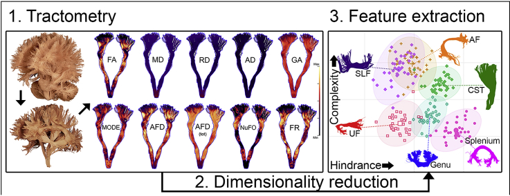
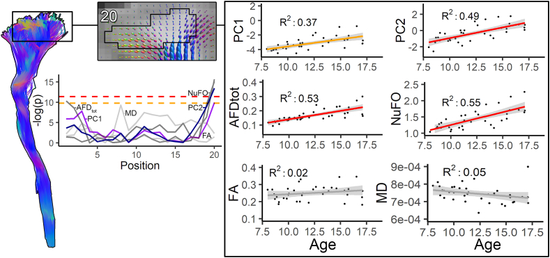
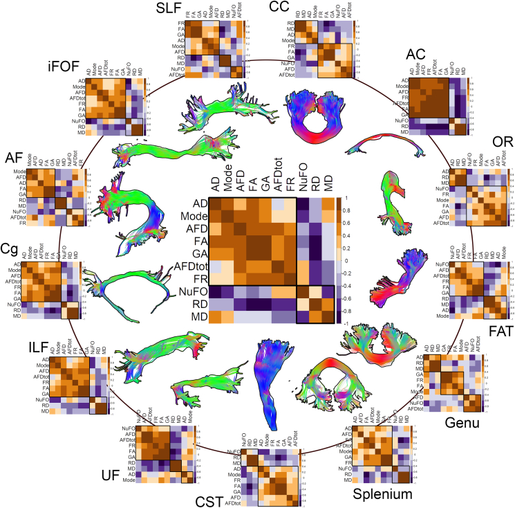
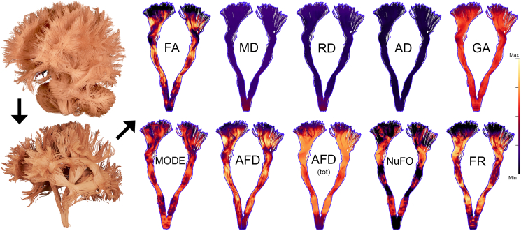
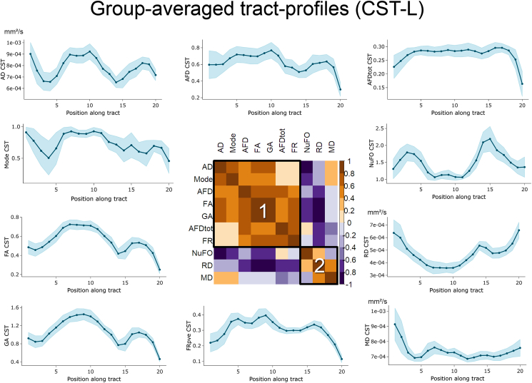
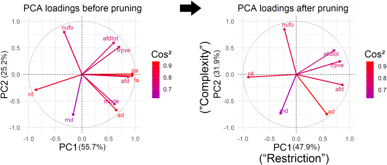
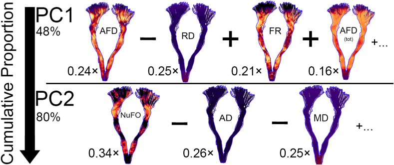
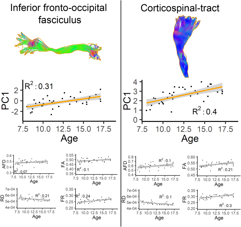
Various diffusion MRI (dMRI) measures have been proposed for characterising tissue microstructure over the last 15 years. Despite the growing number of experiments using different dMRI measures in assessments of white matter, there has been limited work on: 1) examining their covariance along specific pathways; and on 2) combining these different measures to study tissue microstructure. Indeed, it quickly becomes intractable for existing analysis pipelines to process multiple measurements at each voxel and at each vertex forming a streamline, highlighting the need for new ways to visualise or analyse such high-dimensional data. In a sample of 36 typically developing children aged 8-18 years, we profiled various commonly used dMRI measures across 22 brain pathways. Using a data-reduction approach, we identified two biologically-interpretable components that capture 80% of the variance in these dMRI measures. The first derived component captures properties related to hindrance and restriction in tissue microstructure, while the second component reflects characteristics related to tissue complexity and orientational dispersion. We then demonstrate that the components generated by this approach preserve the biological relevance of the original measurements by showing age-related effects across developmentally sensitive pathways. In summary, our findings demonstrate that dMRI analyses can benefit from dimensionality reduction techniques, to help disentangling the neurobiological underpinnings of white matter organisation.
在过去的 15 年中,已经提出了各种扩散磁共振成像(dMRI)测量方法来描述组织微观结构。尽管越来越多的实验使用不同的 dMRI 测量方法来评估白质,但在以下两个方面的工作有限:1) 检查它们在特定路径上的协方差;以及 2) 结合这些不同的测量方法来研究组织微观结构。事实上,现有的分析流水线在每个体素和每个形成流线的顶点处处理多个测量值很快变得难以处理,这突出了需要新的方法来可视化或分析这种高维数据。在一个由 36 名年龄在 8 至 18 岁的典型发育儿童组成的样本中,我们对 22 条大脑通路中的各种常用 dMRI 测量方法进行了分析。我们使用数据减少方法,确定了两个具有生物学解释的分量,这些分量捕获了这些 dMRI 测量值 80%的方差。第一个分量捕获了与组织微观结构中的阻碍和限制相关的特性,而第二个分量反映了与组织复杂性和取向分散相关的特性。然后,我们通过显示与发育敏感通路相关的年龄相关效应,证明了这种方法生成的分量保留了原始测量值的生物学相关性。总之,我们的研究结果表明,dMRI 分析可以从降维技术中受益,以帮助厘清白质组织的神经生物学基础。