Henriques Rafael Neto, Henson Richard, Correia Marta Morgado
Champalimaud Research, Champalimaud Foundation, Lisboa, Portugal.
MRC Cognition and Brain Sciences Unit, University of Cambridge, Cambridge, United Kingdom.
Imaging Neurosci (Camb). 2023 Dec 21;1. doi: 10.1162/imag_a_00051. eCollection 2023.
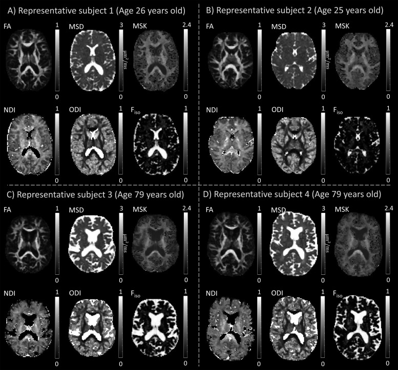
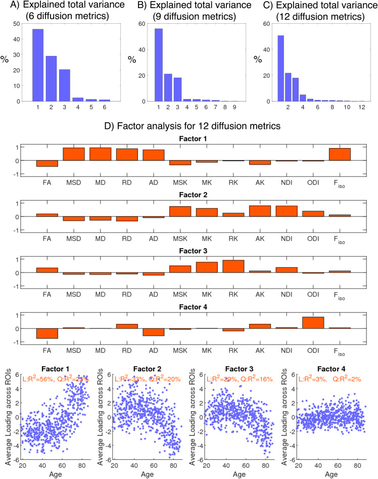
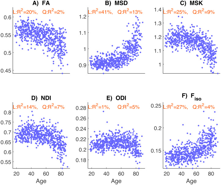
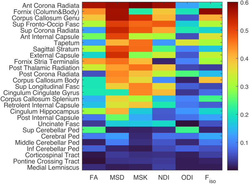
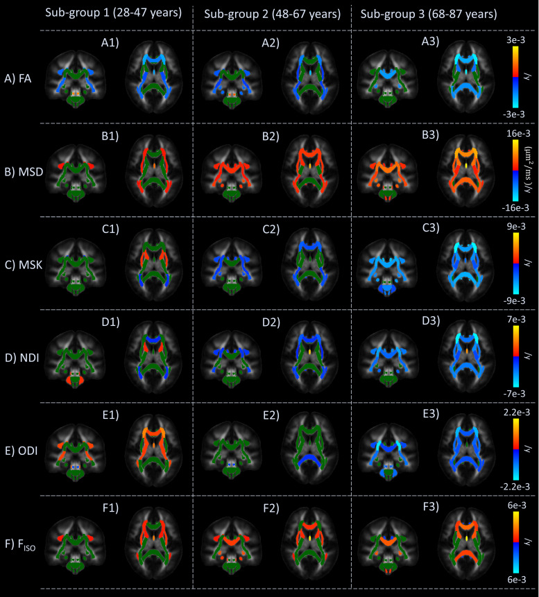
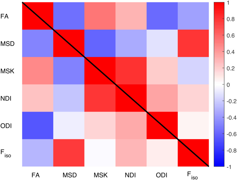
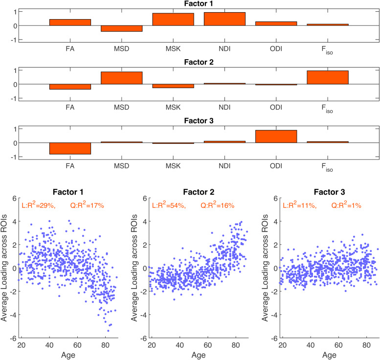
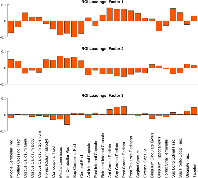
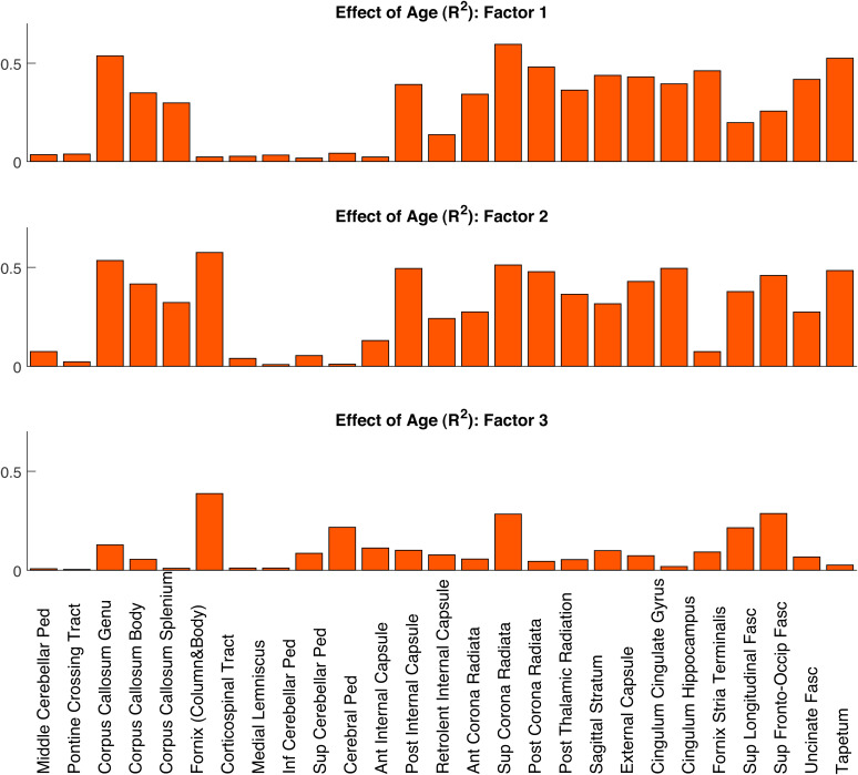
Diffusion Magnetic Resonance Imaging (dMRI) is sensitive to white matter microstructural changes across the human lifespan. Several models have been proposed to provide more sensitive and specific metrics than those provided by the conventional Diffusion Tensor Imaging (DTI) analysis. However, previous results using different metrics have led to contradictory conclusions regarding the effect of age on fibre demyelination and axonal loss in adults. Moreover, it remains unclear whether these metrics provide distinct information about the effects of age, for example, on different white-matter tracts. To address this, we analysed dMRI data from 651 adults approximately uniformly aged from 18 to 88 years in the Cambridge Centre for Ageing and Neuroscience (Cam-CAN) cohort, using six dMRI metrics: Fractional Anisotropy (FA) from standard DTI; Mean Signal Diffusion (MSD) and Mean Signal Kurtosis (MSK) from Diffusional Kurtosis Imaging (DKI) applied to directional averaged diffusion-weighted signals; and Neurite Density Index (NDI), Orientation Dispersion Index (ODI), and isotropic Free water volume fraction (F) estimated from Neurite Orientation Dispersion and Density Imaging (NODDI). Averaging across white-matter regions-of-interest (ROIs), second-order polynomial fits revealed that MSD, MSK, and Fshowed the strongest effects of age, with significant quadratic components suggesting more rapid and sometimes inverted effects in old age. Analysing the data in different age subgroups revealed that some apparent discrepancies in previous studies may be explained by the use of cohorts with different age ranges. Factor analysis of the six metrics across all ROIs revealed three independent factors that can be associated to 1) tissue microscopic properties (e.g., differences in fibre density/myelin), 2) free-water contamination, and 3) tissue configuration complexity (e.g., crossing, dispersing, fanning fibres). While FA captures a combination of different factors, other dMRI metrics are strongly aligned to specific factors (NDI and MSK with Factor 1, Fwith Factor 2, and ODI with Factor 3). To assess whether directional diffusion and kurtosis quantities provide additional information about the effects of age, further factor analyses were also performed, which showed that additional information about the effects of age may be present in radial and axial kurtosis estimates (but not standard axial and radial diffusivity). In summary, our study offers an explanation for previous discrepancies reported in dMRI ageing studies and provides further insights on the interpretation of different dMRI metrics in the context of white-matter microstructural properties.
扩散磁共振成像(dMRI)对人类整个生命周期中的白质微观结构变化敏感。已经提出了几种模型,以提供比传统扩散张量成像(DTI)分析更敏感和特异的指标。然而,先前使用不同指标的结果在年龄对成人纤维脱髓鞘和轴突损失的影响方面得出了相互矛盾的结论。此外,尚不清楚这些指标是否提供了关于年龄影响的不同信息,例如,对不同的白质束的影响。为了解决这个问题,我们分析了来自剑桥衰老与神经科学中心(Cam-CAN)队列中651名年龄在18至88岁之间大致均匀分布的成年人的dMRI数据,使用了六个dMRI指标:标准DTI的分数各向异性(FA);应用于方向平均扩散加权信号的扩散峰度成像(DKI)的平均信号扩散(MSD)和平均信号峰度(MSK);以及从神经突方向离散和密度成像(NODDI)估计的神经突密度指数(NDI)、方向离散指数(ODI)和各向同性自由水体积分数(F)。对感兴趣的白质区域(ROI)进行平均,二阶多项式拟合显示MSD、MSK和F显示出最强的年龄效应,显著的二次成分表明在老年时效应更快,有时甚至相反。对不同年龄亚组的数据进行分析表明,先前研究中一些明显的差异可能是由于使用了不同年龄范围的队列。对所有ROI的六个指标进行因子分析,揭示了三个独立的因子,它们可以与1)组织微观特性(例如,纤维密度/髓磷脂的差异)、2)自由水污染和3)组织构型复杂性(例如,交叉、分散、扇形纤维)相关联。虽然FA捕获了不同因素的组合,但其他dMRI指标与特定因素强烈对齐(NDI和MSK与因子1,F与因子2,ODI与因子3)。为了评估方向扩散和峰度量是否提供了关于年龄影响的额外信息,还进行了进一步的因子分析,结果表明关于年龄影响的额外信息可能存在于径向和轴向峰度估计中(但不是标准的轴向和径向扩散率)。总之,我们的研究为dMRI衰老研究中先前报道的差异提供了解释,并在白质微观结构特性的背景下为不同dMRI指标的解释提供了进一步的见解。