• 文献检索
  • 文档翻译
  • 深度研究
  • 学术资讯
  • Suppr Zotero 插件Zotero 插件
  • 邀请有礼
  • 套餐&价格
  • 历史记录
应用&插件
Suppr Zotero 插件Zotero 插件浏览器插件Mac 客户端Windows 客户端微信小程序
定价
高级版会员购买积分包购买API积分包
服务
文献检索文档翻译深度研究API 文档MCP 服务
关于我们
关于 Suppr公司介绍联系我们用户协议隐私条款
关注我们

Suppr 超能文献

核心技术专利:CN118964589B侵权必究
粤ICP备2023148730 号-1Suppr @ 2026

文献检索

告别复杂PubMed语法,用中文像聊天一样搜索,搜遍4000万医学文献。AI智能推荐,让科研检索更轻松。

立即免费搜索

文件翻译

保留排版,准确专业,支持PDF/Word/PPT等文件格式,支持 12+语言互译。

免费翻译文档

深度研究

AI帮你快速写综述,25分钟生成高质量综述,智能提取关键信息,辅助科研写作。

立即免费体验

深度学习网络在间期和筛查检出癌症的既往阴性乳腺钼靶片中发现了独特的影像学差异:病例对照研究。

Deep learning networks find unique mammographic differences in previous negative mammograms between interval and screen-detected cancers: a case-case study.

机构信息

Department of Bioengineering, University of California-San Francisco Berkeley Joint Program, Room A-C106-B, 1 Irving St, San Francisco, CA, 94143, USA.

Department of Radiology and Biomedical Imaging, UC-San Francisco, San Francisco, CA, 94143, USA.

出版信息

Cancer Imaging. 2019 Jun 22;19(1):41. doi: 10.1186/s40644-019-0227-3.

DOI:10.1186/s40644-019-0227-3
PMID:31228956
原文链接:https://pmc.ncbi.nlm.nih.gov/articles/PMC6589178/
Abstract

BACKGROUND

To determine if mammographic features from deep learning networks can be applied in breast cancer to identify groups at interval invasive cancer risk due to masking beyond using traditional breast density measures.

METHODS

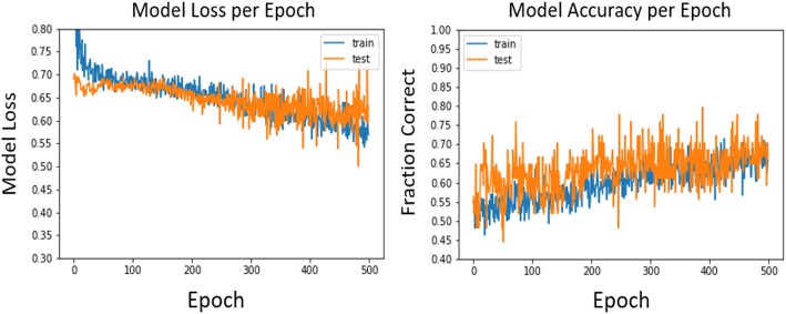
Full-field digital screening mammograms acquired in our clinics between 2006 and 2015 were reviewed. Transfer learning of a deep learning network with weights initialized from ImageNet was performed to classify mammograms that were followed by an invasive interval or screen-detected cancer within 12 months of the mammogram. Hyperparameter optimization was performed and the network was visualized through saliency maps. Prediction loss and accuracy were calculated using this deep learning network. Receiver operating characteristic (ROC) curves and area under the curve (AUC) values were generated with the outcome of interval cancer using the deep learning network and compared to predictions from conditional logistic regression with errors quantified through contingency tables.

RESULTS

Pre-cancer mammograms of 182 interval and 173 screen-detected cancers were split into training/test cases at an 80/20 ratio. Using Breast Imaging-Reporting and Data System (BI-RADS) density alone, the ability to correctly classify interval cancers was moderate (AUC = 0.65). The optimized deep learning model achieved an AUC of 0.82. Contingency table analysis showed the network was correctly classifying 75.2% of the mammograms and that incorrect classifications were slightly more common for the interval cancer mammograms. Saliency maps of each cancer case found that local information could highly drive classification of cases more than global image information.

CONCLUSIONS

Pre-cancerous mammograms contain imaging information beyond breast density that can be identified with deep learning networks to predict the probability of breast cancer detection.

摘要

背景

为了确定深度学习网络的乳腺摄影特征是否可应用于乳腺癌中,以确定因传统乳腺密度测量以外的因素而导致的间隔浸润性癌风险的群体。

方法

回顾了我们诊所于 2006 年至 2015 年间获取的全视野数字化筛查乳腺 X 线照片。通过使用 ImageNet 初始化权重的深度学习网络进行转移学习,以对在乳腺 X 线照片后 12 个月内发生浸润性间隔癌或筛查发现癌症的乳腺 X 线照片进行分类。进行超参数优化,并通过显著图可视化网络。使用此深度学习网络计算预测损失和准确性。使用深度学习网络的间隔癌结果生成接收器工作特征(ROC)曲线和曲线下面积(AUC)值,并与通过误差通过列联表量化的条件逻辑回归的预测进行比较。

结果

将 182 例间隔癌和 173 例筛查发现癌症的癌前乳腺 X 线照片按 80/20 的比例分为训练/测试病例。仅使用乳腺影像报告和数据系统(BI-RADS)密度,正确分类间隔癌的能力为中等(AUC=0.65)。优化后的深度学习模型的 AUC 为 0.82。列联表分析表明,网络正确分类了 75.2%的乳腺 X 线照片,并且间隔癌乳腺 X 线照片的错误分类略为常见。对每个癌症病例的显著图进行分析发现,局部信息可以高度驱动病例的分类,而不是全局图像信息。

结论

癌前乳腺 X 线照片包含除乳腺密度以外的影像学信息,可以通过深度学习网络识别,以预测乳腺癌检测的概率。

https://cdn.ncbi.nlm.nih.gov/pmc/blobs/2a20/6589178/4b141f9ed79e/40644_2019_227_Fig4_HTML.jpg
https://cdn.ncbi.nlm.nih.gov/pmc/blobs/2a20/6589178/757a4033d86a/40644_2019_227_Fig1_HTML.jpg
https://cdn.ncbi.nlm.nih.gov/pmc/blobs/2a20/6589178/9ec0f642581e/40644_2019_227_Fig2_HTML.jpg
https://cdn.ncbi.nlm.nih.gov/pmc/blobs/2a20/6589178/aa3027ffb91a/40644_2019_227_Fig3_HTML.jpg
https://cdn.ncbi.nlm.nih.gov/pmc/blobs/2a20/6589178/4b141f9ed79e/40644_2019_227_Fig4_HTML.jpg

相似文献

1
Deep learning networks find unique mammographic differences in previous negative mammograms between interval and screen-detected cancers: a case-case study.深度学习网络在间期和筛查检出癌症的既往阴性乳腺钼靶片中发现了独特的影像学差异:病例对照研究。
Cancer Imaging. 2019 Jun 22;19(1):41. doi: 10.1186/s40644-019-0227-3.
2
Derived mammographic masking measures based on simulated lesions predict the risk of interval cancer after controlling for known risk factors: a case-case analysis.基于模拟病变的衍生乳腺 X 线摄影屏蔽措施可预测已知危险因素控制后间隔期癌症的风险:病例对照分析。
Med Phys. 2019 Mar;46(3):1309-1316. doi: 10.1002/mp.13410. Epub 2019 Feb 14.
3
Novel mammographic image features differentiate between interval and screen-detected breast cancer: a case-case study.新型乳腺钼靶图像特征可区分间期癌和筛查发现的乳腺癌:一项病例对照研究。
Breast Cancer Res. 2016 Oct 5;18(1):100. doi: 10.1186/s13058-016-0761-x.
4
Deep Learning Predicts Interval and Screening-detected Cancer from Screening Mammograms: A Case-Case-Control Study in 6369 Women.深度学习预测筛查性乳房 X 光照片中的间隔期和筛查检出性癌症:6369 名女性的病例对照研究。
Radiology. 2021 Dec;301(3):550-558. doi: 10.1148/radiol.2021203758. Epub 2021 Sep 7.
5
Automated pectoral muscle identification on MLO-view mammograms: Comparison of deep neural network to conventional computer vision.基于 MLO 视图的乳腺钼靶片中自动胸大肌识别:深度神经网络与传统计算机视觉的比较。
Med Phys. 2019 May;46(5):2103-2114. doi: 10.1002/mp.13451. Epub 2019 Mar 12.
6
Predicting interval and screen-detected breast cancers from mammographic density defined by different brightness thresholds.根据不同亮度阈值定义的乳腺密度预测间期和筛查性乳腺癌。
Breast Cancer Res. 2018 Dec 13;20(1):152. doi: 10.1186/s13058-018-1081-0.
7
Mass detection in digital breast tomosynthesis: Deep convolutional neural network with transfer learning from mammography.数字乳腺断层合成中的肿块检测:基于乳腺X线摄影迁移学习的深度卷积神经网络
Med Phys. 2016 Dec;43(12):6654. doi: 10.1118/1.4967345.
8
Mammographic density and structural features can individually and jointly contribute to breast cancer risk assessment in mammography screening: a case-control study.乳腺X线密度和结构特征在乳腺X线筛查中可单独或共同影响乳腺癌风险评估:一项病例对照研究。
BMC Cancer. 2016 Jul 7;16:414. doi: 10.1186/s12885-016-2450-7.
9
Understanding Clinical Mammographic Breast Density Assessment: a Deep Learning Perspective.理解临床乳腺 X 线摄影乳房密度评估:深度学习视角。
J Digit Imaging. 2018 Aug;31(4):387-392. doi: 10.1007/s10278-017-0022-2.
10
Predicting Breast Cancer by Applying Deep Learning to Linked Health Records and Mammograms.应用深度学习对健康记录和乳腺 X 光照片进行联合分析,以预测乳腺癌。
Radiology. 2019 Aug;292(2):331-342. doi: 10.1148/radiol.2019182622. Epub 2019 Jun 18.

引用本文的文献

1
Current Strategies to Reducing Interval Breast Cancers: A Systematic Review.降低间期乳腺癌的当前策略:一项系统综述
Breast Cancer (Dove Med Press). 2025 Jun 27;17:531-544. doi: 10.2147/BCTT.S532884. eCollection 2025.
2
The Future of Breast Cancer Organized Screening Program Through Artificial Intelligence: A Scoping Review.通过人工智能开展乳腺癌组织筛查项目的未来:一项范围综述
Healthcare (Basel). 2025 Feb 10;13(4):378. doi: 10.3390/healthcare13040378.
3
CSA-Net: Channel and Spatial Attention-Based Network for Mammogram and Ultrasound Image Classification.

本文引用的文献

1
Diffusion tensor imaging parameters in differentiation recurrent breast cancer from post-operative changes in patients with breast-conserving surgery.弥散张量成像参数在鉴别保乳手术后患者复发性乳腺癌与术后改变中的作用。
Eur J Radiol. 2019 Feb;111:76-80. doi: 10.1016/j.ejrad.2018.12.022. Epub 2018 Dec 30.
2
A Deep Learning Model to Predict a Diagnosis of Alzheimer Disease by Using F-FDG PET of the Brain.利用大脑 F-FDG PET 预测阿尔茨海默病诊断的深度学习模型。
Radiology. 2019 Feb;290(2):456-464. doi: 10.1148/radiol.2018180958. Epub 2018 Nov 6.
3
Mammographic Breast Density Assessment Using Deep Learning: Clinical Implementation.
CSA-Net:用于乳房X光片和超声图像分类的基于通道和空间注意力的网络
J Imaging. 2024 Oct 16;10(10):256. doi: 10.3390/jimaging10100256.
4
Artificial Intelligence-Driven Mammography-Based Future Breast Cancer Risk Prediction: A Systematic Review.基于人工智能驱动的乳腺钼靶摄影的未来乳腺癌风险预测:一项系统综述。
J Am Coll Radiol. 2024 Feb;21(2):319-328. doi: 10.1016/j.jacr.2023.10.018. Epub 2023 Nov 8.
5
Improving the malignancy prediction of breast cancer based on the integration of radiomics features from dual-view mammography and clinical parameters.基于双视图乳腺 X 线摄影和临床参数的放射组学特征融合提高乳腺癌恶性程度预测能力。
Clin Exp Med. 2023 Oct;23(6):2357-2368. doi: 10.1007/s10238-022-00944-8. Epub 2022 Nov 21.
6
Artificial intelligence in mammographic phenotyping of breast cancer risk: a narrative review.人工智能在乳腺癌风险的乳腺摄影表型中的应用:叙述性综述。
Breast Cancer Res. 2022 Feb 20;24(1):14. doi: 10.1186/s13058-022-01509-z.
7
An Open-Source Framework for Automated High-Throughput Cell Biology Experiments.用于自动化高通量细胞生物学实验的开源框架。
Front Cell Dev Biol. 2021 Sep 24;9:697584. doi: 10.3389/fcell.2021.697584. eCollection 2021.
8
Deep Learning Predicts Interval and Screening-detected Cancer from Screening Mammograms: A Case-Case-Control Study in 6369 Women.深度学习预测筛查性乳房 X 光照片中的间隔期和筛查检出性癌症:6369 名女性的病例对照研究。
Radiology. 2021 Dec;301(3):550-558. doi: 10.1148/radiol.2021203758. Epub 2021 Sep 7.
基于深度学习的乳腺 X 线摄影密度评估:临床应用。
Radiology. 2019 Jan;290(1):52-58. doi: 10.1148/radiol.2018180694. Epub 2018 Oct 16.
4
Deep Learning to Distinguish Recalled but Benign Mammography Images in Breast Cancer Screening.深度学习在乳腺癌筛查中区分召回的良性乳腺 X 光图像。
Clin Cancer Res. 2018 Dec 1;24(23):5902-5909. doi: 10.1158/1078-0432.CCR-18-1115. Epub 2018 Oct 11.
5
Automated and Clinical Breast Imaging Reporting and Data System Density Measures Predict Risk for Screen-Detected and Interval Cancers: A Case-Control Study.自动化和临床乳腺成像报告和数据系统密度测量预测筛查和间期癌症的风险:病例对照研究。
Ann Intern Med. 2018 Jun 5;168(11):757-765. doi: 10.7326/M17-3008. Epub 2018 May 1.
6
Deep learning beyond cats and dogs: recent advances in diagnosing breast cancer with deep neural networks.超越猫狗识别的深度学习:深度神经网络在乳腺癌诊断中的最新进展
Br J Radiol. 2018 Sep;91(1089):20170545. doi: 10.1259/bjr.20170545. Epub 2018 Apr 10.
7
Detecting and classifying lesions in mammograms with Deep Learning.深度学习在乳腺 X 光片中检测和分类病灶。
Sci Rep. 2018 Mar 15;8(1):4165. doi: 10.1038/s41598-018-22437-z.
8
A survey on deep learning in medical image analysis.深度学习在医学图像分析中的应用研究综述。
Med Image Anal. 2017 Dec;42:60-88. doi: 10.1016/j.media.2017.07.005. Epub 2017 Jul 26.
9
Mammographic texture and risk of breast cancer by tumor type and estrogen receptor status.乳腺钼靶纹理与不同肿瘤类型及雌激素受体状态的乳腺癌风险
Breast Cancer Res. 2016 Dec 6;18(1):122. doi: 10.1186/s13058-016-0778-1.
10
National Performance Benchmarks for Modern Screening Digital Mammography: Update from the Breast Cancer Surveillance Consortium.现代筛查数字化乳腺摄影的国家性能基准:来自乳腺癌监测联盟的更新
Radiology. 2017 Apr;283(1):49-58. doi: 10.1148/radiol.2016161174. Epub 2016 Dec 5.