• 文献检索
  • 文档翻译
  • 深度研究
  • 学术资讯
  • Suppr Zotero 插件Zotero 插件
  • 邀请有礼
  • 套餐&价格
  • 历史记录
应用&插件
Suppr Zotero 插件Zotero 插件浏览器插件Mac 客户端Windows 客户端微信小程序
定价
高级版会员购买积分包购买API积分包
服务
文献检索文档翻译深度研究API 文档MCP 服务
关于我们
关于 Suppr公司介绍联系我们用户协议隐私条款
关注我们

Suppr 超能文献

核心技术专利:CN118964589B侵权必究
粤ICP备2023148730 号-1Suppr @ 2026

文献检索

告别复杂PubMed语法,用中文像聊天一样搜索,搜遍4000万医学文献。AI智能推荐,让科研检索更轻松。

立即免费搜索

文件翻译

保留排版,准确专业,支持PDF/Word/PPT等文件格式,支持 12+语言互译。

免费翻译文档

深度研究

AI帮你快速写综述,25分钟生成高质量综述,智能提取关键信息,辅助科研写作。

立即免费体验

基于机器学习,利用CT纹理特征鉴别胰腺浆液性囊腺瘤与黏液性囊腺瘤

Discrimination of Pancreatic Serous Cystadenomas From Mucinous Cystadenomas With CT Textural Features: Based on Machine Learning.

作者信息

Yang Jing, Guo Xinli, Ou Xuejin, Zhang Weiwei, Ma Xuelei

机构信息

State Key Laboratory of Biotherapy, Department of Biotherapy, West China Hospital, Cancer Center, Sichuan University, Chengdu, China.

West China Hospital, West China School of Medicine, Sichuan University, Chengdu, China.

出版信息

Front Oncol. 2019 Jun 12;9:494. doi: 10.3389/fonc.2019.00494. eCollection 2019.

DOI:10.3389/fonc.2019.00494
PMID:31245294
原文链接:https://pmc.ncbi.nlm.nih.gov/articles/PMC6581751/
Abstract

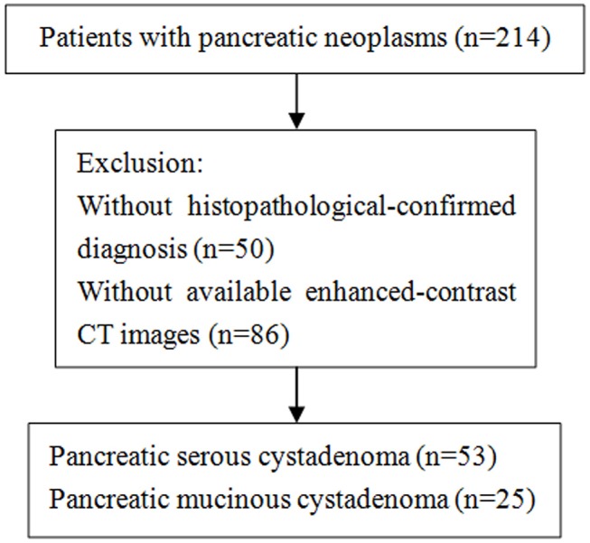
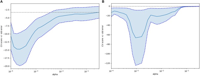
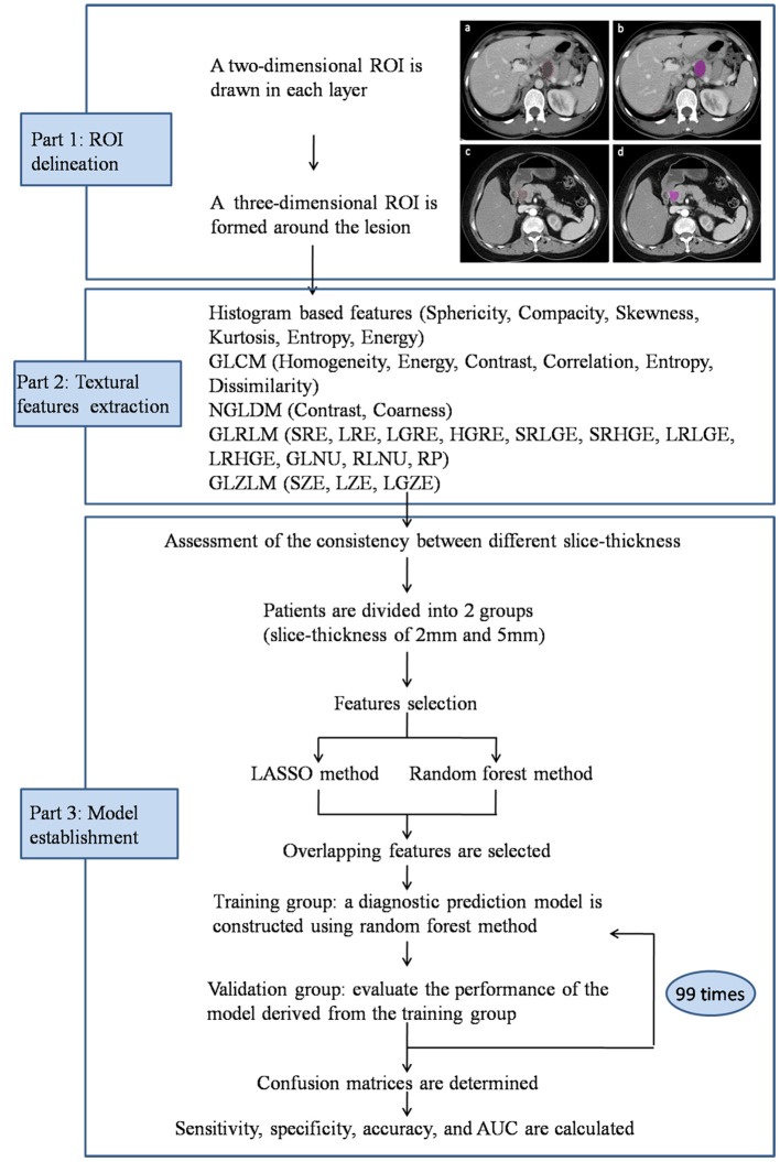
This study was designed to estimate the performance of textural features derived from contrast-enhanced CT in the differential diagnosis of pancreatic serous cystadenomas and pancreatic mucinous cystadenomas. Fifty-three patients with pancreatic serous cystadenoma and 25 patients with pancreatic mucinous cystadenoma were included. Textural parameters of the pancreatic neoplasms were extracted using the LIFEx software, and were analyzed using random forest and Least Absolute Shrinkage and Selection Operator (LASSO) methods. Patients were randomly divided into training and validation sets with a ratio of 4:1; random forest method was adopted to constructed a diagnostic prediction model. Scoring metrics included sensitivity, specificity, accuracy, and AUC. Radiomics features extracted from contrast-enhanced CT were able to discriminate pancreatic mucinous cystadenomas from serous cystadenomas in both the training group (slice thickness of 2 mm, AUC 0.77, sensitivity 0.95, specificity 0.83, accuracy 0.85; slice thickness of 5 mm, AUC 0.72, sensitivity 0.90, specificity 0.84, accuracy 0.86) and the validation group (slice thickness of 2 mm, AUC 0.66, sensitivity 0.86, specificity 0.71, accuracy 0.74; slice thickness of 5 mm, AUC 0.75, sensitivity 0.85, specificity 0.83, accuracy 0.83). In conclusion, our study provided preliminary evidence that textural features derived from CT images were useful in differential diagnosis of pancreatic mucinous cystadenomas and serous cystadenomas, which may provide a non-invasive approach to determine whether surgery is needed in clinical practice. However, multicentre studies with larger sample size are needed to confirm these results.

摘要

本研究旨在评估对比增强CT衍生的纹理特征在胰腺浆液性囊腺瘤和胰腺黏液性囊腺瘤鉴别诊断中的性能。纳入了53例胰腺浆液性囊腺瘤患者和25例胰腺黏液性囊腺瘤患者。使用LIFEx软件提取胰腺肿瘤的纹理参数,并采用随机森林和最小绝对收缩和选择算子(LASSO)方法进行分析。患者以4:1的比例随机分为训练集和验证集;采用随机森林方法构建诊断预测模型。评分指标包括敏感性、特异性、准确性和AUC。在训练组(层厚2 mm,AUC 0.77,敏感性0.95,特异性0.83,准确性0.85;层厚5 mm,AUC 0.72,敏感性0.90,特异性0.84,准确性0.86)和验证组(层厚2 mm,AUC 0.66,敏感性0.86,特异性0.71,准确性0.74;层厚5 mm,AUC 0.75,敏感性0.85,特异性0.83,准确性0.83)中,对比增强CT提取的影像组学特征均能够区分胰腺黏液性囊腺瘤和浆液性囊腺瘤。总之,我们的研究提供了初步证据,表明CT图像衍生的纹理特征在胰腺黏液性囊腺瘤和浆液性囊腺瘤的鉴别诊断中有用,这可能为临床实践中确定是否需要手术提供一种非侵入性方法。然而,需要更大样本量的多中心研究来证实这些结果。

https://cdn.ncbi.nlm.nih.gov/pmc/blobs/5a41/6581751/de07e9b05f23/fonc-09-00494-g0003.jpg
https://cdn.ncbi.nlm.nih.gov/pmc/blobs/5a41/6581751/4feefe9821e1/fonc-09-00494-g0001.jpg
https://cdn.ncbi.nlm.nih.gov/pmc/blobs/5a41/6581751/c8341fa440c4/fonc-09-00494-g0002.jpg
https://cdn.ncbi.nlm.nih.gov/pmc/blobs/5a41/6581751/de07e9b05f23/fonc-09-00494-g0003.jpg
https://cdn.ncbi.nlm.nih.gov/pmc/blobs/5a41/6581751/4feefe9821e1/fonc-09-00494-g0001.jpg
https://cdn.ncbi.nlm.nih.gov/pmc/blobs/5a41/6581751/c8341fa440c4/fonc-09-00494-g0002.jpg
https://cdn.ncbi.nlm.nih.gov/pmc/blobs/5a41/6581751/de07e9b05f23/fonc-09-00494-g0003.jpg

相似文献

1
Discrimination of Pancreatic Serous Cystadenomas From Mucinous Cystadenomas With CT Textural Features: Based on Machine Learning.基于机器学习,利用CT纹理特征鉴别胰腺浆液性囊腺瘤与黏液性囊腺瘤
Front Oncol. 2019 Jun 12;9:494. doi: 10.3389/fonc.2019.00494. eCollection 2019.
2
Differential diagnosis of pancreatic serous cystadenoma and mucinous cystadenoma: utility of textural features in combination with morphological characteristics.胰腺浆液性囊腺瘤和黏液性囊腺瘤的鉴别诊断:纹理特征结合形态学特征的应用。
BMC Cancer. 2019 Dec 16;19(1):1223. doi: 10.1186/s12885-019-6421-7.
3
[Characterization and diagnostic accuracy of serous cystadenomas and mucinous neoplasms of the pancreas with multi-slice helical computed tomography].[多层螺旋计算机断层扫描对胰腺浆液性囊腺瘤和黏液性肿瘤的特征分析及诊断准确性]
Zhongguo Yi Xue Ke Xue Yuan Xue Bao. 2007 Apr;29(2):232-7.
4
Pancreatic Serous Cystic Neoplasms and Mucinous Cystic Neoplasms: Differential Diagnosis by Combining Imaging Features and Enhanced CT Texture Analysis.胰腺浆液性囊性肿瘤和黏液性囊性肿瘤:结合影像特征与增强CT纹理分析进行鉴别诊断
Front Oncol. 2021 Dec 23;11:745001. doi: 10.3389/fonc.2021.745001. eCollection 2021.
5
[Multi-slice computed tomography for diagnosis of mucinous cystic neoplasms and serous cystadenomas of the pancreas].多层螺旋计算机断层扫描在胰腺黏液性囊性肿瘤和浆液性囊腺瘤诊断中的应用
Nan Fang Yi Ke Da Xue Xue Bao. 2012 Jan;32(1):19-22.
6
Macrocystic pancreatic cystadenoma: The role of EUS and cyst fluid analysis in distinguishing mucinous and serous lesions.大囊型胰腺囊腺瘤:超声内镜及囊液分析在鉴别黏液性和浆液性病变中的作用
Gastrointest Endosc. 2004 Jun;59(7):823-9. doi: 10.1016/s0016-5107(04)00346-3.
7
Application of CT-Based Radiomics in Discriminating Pancreatic Cystadenomas From Pancreatic Neuroendocrine Tumors Using Machine Learning Methods.基于CT的影像组学在使用机器学习方法鉴别胰腺囊腺瘤与胰腺神经内分泌肿瘤中的应用
Front Oncol. 2021 Jul 22;11:606677. doi: 10.3389/fonc.2021.606677. eCollection 2021.
8
Cyst fluid analysis in the differential diagnosis of pancreatic cysts. A comparison of pseudocysts, serous cystadenomas, mucinous cystic neoplasms, and mucinous cystadenocarcinoma.胰腺囊肿鉴别诊断中的囊液分析。假性囊肿、浆液性囊腺瘤、黏液性囊性肿瘤及黏液性囊腺癌的比较。
Ann Surg. 1993 Jan;217(1):41-7. doi: 10.1097/00000658-199301000-00008.
9
Preoperative differentiation of pancreatic mucinous cystic neoplasm from macrocystic serous cystic adenoma using radiomics: Preliminary findings and comparison with radiological model.基于影像组学的术前鉴别胰腺黏液性囊性肿瘤与大囊型浆液性囊腺瘤:初步研究结果与影像学模型比较。
Eur J Radiol. 2020 Jan;122:108747. doi: 10.1016/j.ejrad.2019.108747. Epub 2019 Nov 14.
10
CT-Based Radiomics Analysis for Preoperative Diagnosis of Pancreatic Mucinous Cystic Neoplasm and Atypical Serous Cystadenomas.基于CT的放射组学分析在胰腺黏液性囊性肿瘤和非典型浆液性囊腺瘤术前诊断中的应用
Front Oncol. 2021 Jun 11;11:621520. doi: 10.3389/fonc.2021.621520. eCollection 2021.

引用本文的文献

1
Advancements in Radiomics-Based AI for Pancreatic Ductal Adenocarcinoma.基于放射组学的人工智能在胰腺导管腺癌中的进展。
Bioengineering (Basel). 2025 Aug 6;12(8):849. doi: 10.3390/bioengineering12080849.
2
Application of Artificial Intelligence in Pancreatic Cyst Management: A Systematic Review.人工智能在胰腺囊肿管理中的应用:一项系统综述
Cancers (Basel). 2025 Aug 2;17(15):2558. doi: 10.3390/cancers17152558.
3
Evolving and Novel Applications of Artificial Intelligence in Cancer Imaging.人工智能在癌症成像中的不断发展与新应用

本文引用的文献

1
Two-dimensional Texture Analysis Based on CT Images to Differentiate Pancreatic Lymphoma and Pancreatic Adenocarcinoma: A Preliminary Study.基于 CT 图像的二维纹理分析鉴别胰腺淋巴瘤与胰腺腺癌:一项初步研究。
Acad Radiol. 2019 Aug;26(8):e189-e195. doi: 10.1016/j.acra.2018.07.021. Epub 2018 Sep 5.
2
LIFEx: A Freeware for Radiomic Feature Calculation in Multimodality Imaging to Accelerate Advances in the Characterization of Tumor Heterogeneity.LIFEx:一种用于多模态成像中放射组学特征计算的免费软件,可加速肿瘤异质性特征描述的进展。
Cancer Res. 2018 Aug 15;78(16):4786-4789. doi: 10.1158/0008-5472.CAN-18-0125. Epub 2018 Jun 29.
3
Cancers (Basel). 2025 Apr 30;17(9):1510. doi: 10.3390/cancers17091510.
4
Machine Learning-Driven Radiomics Analysis for Distinguishing Mucinous and Non-Mucinous Pancreatic Cystic Lesions: A Multicentric Study.机器学习驱动的放射组学分析用于鉴别胰腺黏液性和非黏液性囊性病变:一项多中心研究
J Imaging. 2025 Feb 20;11(3):68. doi: 10.3390/jimaging11030068.
5
Artificial Intelligence in Pancreatic Imaging: A Systematic Review.胰腺成像中的人工智能:一项系统综述。
United European Gastroenterol J. 2025 Feb;13(1):55-77. doi: 10.1002/ueg2.12723. Epub 2025 Jan 26.
6
Radiomics advances in the evaluation of pancreatic cystic neoplasms.胰腺囊性肿瘤评估中的影像组学进展。
Heliyon. 2024 Jan 30;10(3):e25535. doi: 10.1016/j.heliyon.2024.e25535. eCollection 2024 Feb 15.
7
A novel CT-based radiomics model for predicting response and prognosis of chemoradiotherapy in esophageal squamous cell carcinoma.一种基于 CT 的新型放射组学模型,用于预测食管鳞癌放化疗反应和预后。
Sci Rep. 2024 Jan 23;14(1):2039. doi: 10.1038/s41598-024-52418-4.
8
Serous Cystadenoma: A Review on Diagnosis and Management.浆液性囊腺瘤:诊断与治疗综述
J Clin Med. 2023 Nov 25;12(23):7306. doi: 10.3390/jcm12237306.
9
Risk Assessment and Pancreatic Cancer: Diagnostic Management and Artificial Intelligence.风险评估与胰腺癌:诊断管理与人工智能
Cancers (Basel). 2023 Jan 5;15(2):351. doi: 10.3390/cancers15020351.
10
The Convergence of FTIR and EVs: Emergence Strategy for Non-Invasive Cancer Markers Discovery.傅里叶变换红外光谱(FTIR)与细胞外囊泡(EVs)的融合:发现非侵入性癌症标志物的新策略
Diagnostics (Basel). 2022 Dec 21;13(1):22. doi: 10.3390/diagnostics13010022.
Primary central nervous system lymphoma and atypical glioblastoma: Differentiation using radiomics approach.
原发性中枢神经系统淋巴瘤和非典型性脑胶质瘤:基于放射组学方法的鉴别诊断。
Eur Radiol. 2018 Sep;28(9):3832-3839. doi: 10.1007/s00330-018-5368-4. Epub 2018 Apr 6.
4
Ability of FDG PET and CT radiomics features to differentiate between primary and metastatic lung lesions.FDG PET 和 CT 影像组学特征区分原发性和转移性肺病变的能力。
Eur J Nucl Med Mol Imaging. 2018 Sep;45(10):1649-1660. doi: 10.1007/s00259-018-3987-2. Epub 2018 Apr 6.
5
Diagnostic Performance of Mammographic Texture Analysis in the Differential Diagnosis of Benign and Malignant Breast Tumors.乳腺摄影纹理分析在鉴别良恶性乳腺肿瘤中的诊断性能。
Clin Breast Cancer. 2018 Aug;18(4):e621-e627. doi: 10.1016/j.clbc.2017.11.004. Epub 2017 Nov 9.
6
Circulating tumour DNA methylation markers for diagnosis and prognosis of hepatocellular carcinoma.循环肿瘤 DNA 甲基化标志物用于肝细胞癌的诊断和预后。
Nat Mater. 2017 Nov;16(11):1155-1161. doi: 10.1038/nmat4997. Epub 2017 Oct 9.
7
CT Texture Analysis Potentially Predicts Local Failure in Head and Neck Squamous Cell Carcinoma Treated with Chemoradiotherapy.CT 纹理分析可能预测头颈部鳞状细胞癌经放化疗后局部失败。
AJNR Am J Neuroradiol. 2017 Dec;38(12):2334-2340. doi: 10.3174/ajnr.A5407. Epub 2017 Oct 12.
8
Endometrial Carcinoma: MR Imaging-based Texture Model for Preoperative Risk Stratification-A Preliminary Analysis.子宫内膜癌:基于磁共振成像的纹理模型用于术前风险分层——初步分析。
Radiology. 2017 Sep;284(3):748-757. doi: 10.1148/radiol.2017161950. Epub 2017 May 10.
9
CT-based texture analysis potentially provides prognostic information complementary to interim fdg-pet for patients with hodgkin's and aggressive non-hodgkin's lymphomas.基于CT的纹理分析可能为霍奇金淋巴瘤和侵袭性非霍奇金淋巴瘤患者提供与中期氟代脱氧葡萄糖正电子发射断层扫描(FDG-PET)互补的预后信息。
Eur Radiol. 2017 Mar;27(3):1012-1020. doi: 10.1007/s00330-016-4470-8. Epub 2016 Jul 5.
10
Pancreatic Cystic Neoplasms: An Update.胰腺囊性肿瘤:最新进展
Gastroenterol Clin North Am. 2016 Mar;45(1):67-81. doi: 10.1016/j.gtc.2015.10.006. Epub 2016 Jan 13.