Saxon Incubator for Clinical Translation (SIKT), University of Leipzig, Leipzig, Germany.
Faculty of Veterinary Medicine, Equine Clinic & Hospital, University of Leipzig, Leipzig, Germany.
PLoS One. 2019 Jun 27;14(6):e0218949. doi: 10.1371/journal.pone.0218949. eCollection 2019.
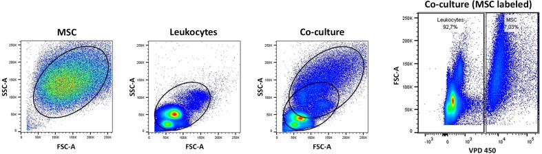
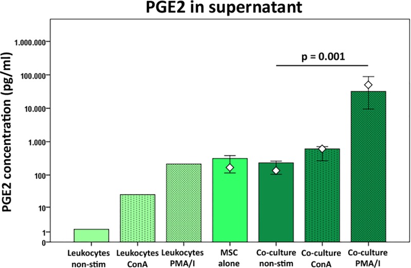
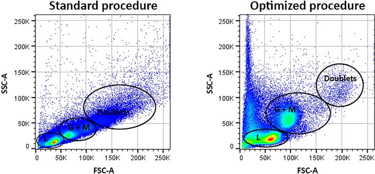
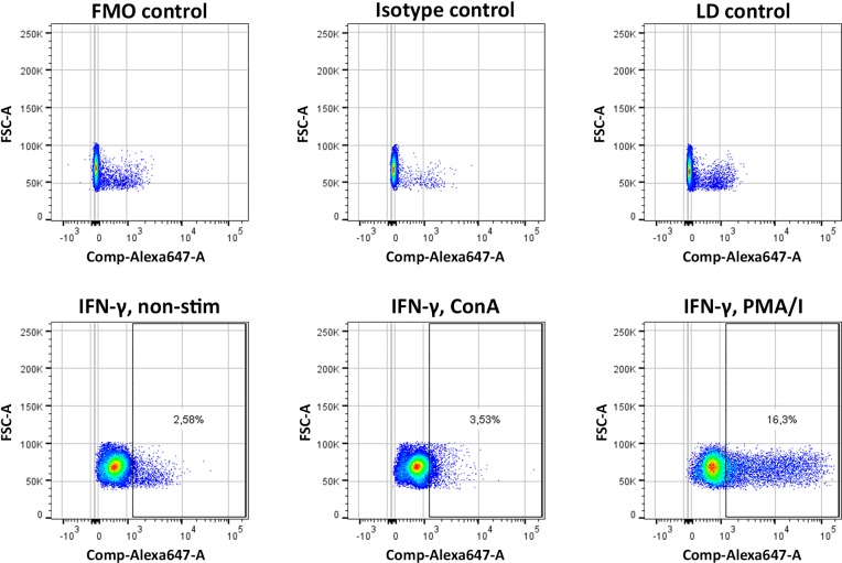
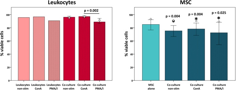
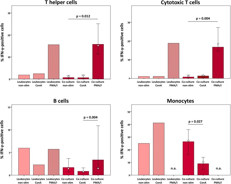
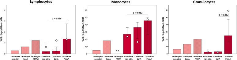
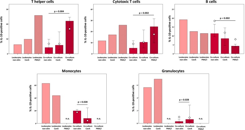
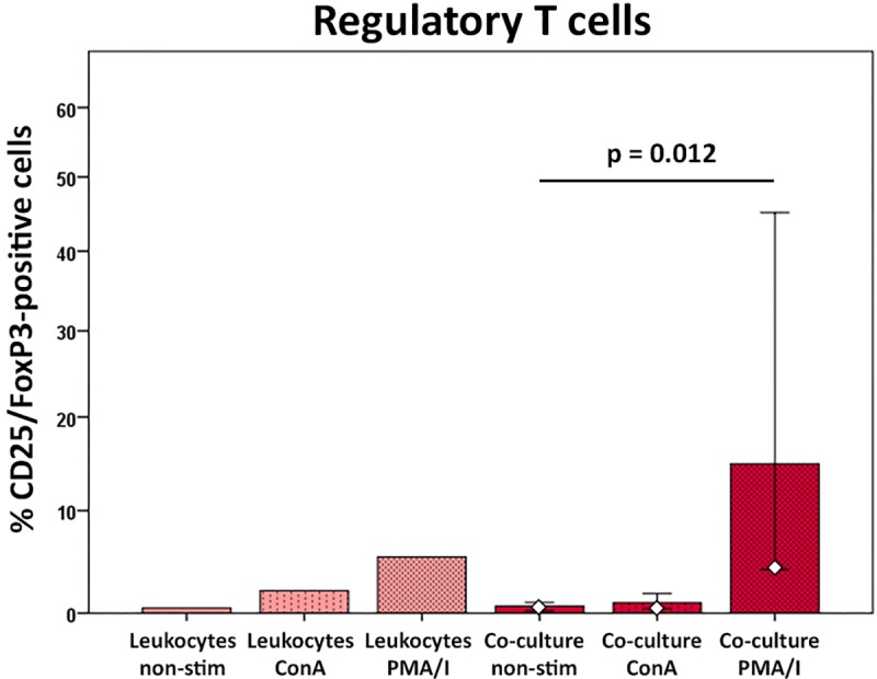
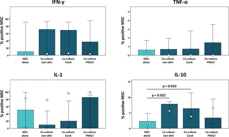
The immunomodulatory potential of multipotent mesenchymal stromal cells (MSC) provides a basis for current and future regenerative therapies. In this study, we established an approach that allows to address the effects of pro-inflammatory stimulation and co-culture with MSC on different specific leukocyte subpopulations. Equine peripheral blood leukocyte recovery was optimized to preserve all leukocyte subpopulations and leukocyte activation regimes were evaluated. Allogeneic labeled equine adipose-derived MSC were then subjected to direct co-culture with either non-stimulated, concanavalin A (ConA)-activated or phosphate 12-myristate 13-acetate and ionomycin (PMA/I)-activated leukocytes. Subsequently, production of the cytokines interferon-γ (IFN- γ), interleukin-1 (IL-1) and tumor necrosis factor-α (TNF-α) and presence of FoxP3 were determined in specific cell populations using multicolor flow cytometry. Prostaglandin E2 (PGE2) was measured in the supernatants. ConA-stimulation induced mild activation of leukocytes, whereas PMA/I-stimulation led to strong activation. In T cells, PMA/I promoted production of all cytokines, with no distinct suppressive effects of MSC. However, increased numbers of CD25/FoxP3-positive cells indicated that MSC supported regulatory T cell differentiation in PMA/I-activated leukocyte cultures. MSC also reduced numbers of cytokine-producing B cells and granulocytes, mostly irrespective of preceding leukocyte activation, and reversed the stimulatory effect of ConA on IFN-γ production in monocytes. Illustrating the possible suppressive mechanisms, higher numbers of MSC produced IL-10 when co-cultured with non-stimulated or ConA-activated leukocytes. This was not observed in co-culture with PMA/I-activated leukocytes. However, PGE2 concentration in the supernatant was highest in the co-culture with PMA/I-activated leukocytes, suggesting that PGE2 could still mediate modulatory effects in strongly inflammatory environment. These context- and cell type-specific modulatory effects observed give insight into the interactions between MSC and different types of immune cells and highlight the roles of IL-10 and PGE2 in MSC-mediated immunomodulation. The approach presented could provide a basis for further functional MSC characterization and the development of potency assays.
多能间充质基质细胞(MSC)的免疫调节潜能为当前和未来的再生治疗提供了基础。在这项研究中,我们建立了一种方法,允许研究促炎刺激和与 MSC 共培养对不同特定白细胞亚群的影响。优化了马外周血白细胞回收以保留所有白细胞亚群,并评估了白细胞激活方案。然后,将同种异体标记的马脂肪来源的 MSC 直接与未经刺激、刀豆蛋白 A(ConA)激活或磷酸 12-肉豆蔻酸 13-乙酸盐和离子霉素(PMA/I)激活的白细胞共培养。随后,使用多色流式细胞术在特定细胞群中确定细胞因子干扰素-γ(IFN-γ)、白细胞介素-1(IL-1)和肿瘤坏死因子-α(TNF-α)的产生和 FoxP3 的存在。在培养物上清液中测量前列腺素 E2(PGE2)。ConA 刺激导致白细胞轻度激活,而 PMA/I 刺激导致强烈激活。在 T 细胞中,PMA/I 促进所有细胞因子的产生,MSC 没有明显的抑制作用。然而,CD25/FoxP3 阳性细胞数量的增加表明 MSC 支持 PMA/I 激活的白细胞培养物中调节性 T 细胞的分化。MSC 还减少了产生细胞因子的 B 细胞和粒细胞的数量,这主要与先前的白细胞激活无关,并逆转了 ConA 对单核细胞 IFN-γ产生的刺激作用。说明可能的抑制机制,当与未刺激或 ConA 激活的白细胞共培养时,MSC 产生更多的 IL-10。在与 PMA/I 激活的白细胞共培养时未观察到这种情况。然而,在与 PMA/I 激活的白细胞共培养物中,上清液中 PGE2 的浓度最高,这表明 PGE2 仍可在强烈炎症环境中发挥调节作用。观察到的这种与上下文和细胞类型相关的调节作用深入了解了 MSC 与不同类型免疫细胞之间的相互作用,并强调了 IL-10 和 PGE2 在 MSC 介导的免疫调节中的作用。所提出的方法可以为进一步的 MSC 功能表征和效力测定的发展提供基础。