• 文献检索
  • 文档翻译
  • 深度研究
  • 学术资讯
  • Suppr Zotero 插件Zotero 插件
  • 邀请有礼
  • 套餐&价格
  • 历史记录
应用&插件
Suppr Zotero 插件Zotero 插件浏览器插件Mac 客户端Windows 客户端微信小程序
定价
高级版会员购买积分包购买API积分包
服务
文献检索文档翻译深度研究API 文档MCP 服务
关于我们
关于 Suppr公司介绍联系我们用户协议隐私条款
关注我们

Suppr 超能文献

核心技术专利:CN118964589B侵权必究
粤ICP备2023148730 号-1Suppr @ 2026

文献检索

告别复杂PubMed语法,用中文像聊天一样搜索,搜遍4000万医学文献。AI智能推荐,让科研检索更轻松。

立即免费搜索

文件翻译

保留排版,准确专业,支持PDF/Word/PPT等文件格式,支持 12+语言互译。

免费翻译文档

深度研究

AI帮你快速写综述,25分钟生成高质量综述,智能提取关键信息,辅助科研写作。

立即免费体验

肌动蛋白清除促进免疫突触极化的动力蛋白积累。

Actin clearance promotes polarized dynein accumulation at the immunological synapse.

机构信息

Immunology Program, Memorial Sloan-Kettering Cancer Center, New York, NY, United States of America.

出版信息

PLoS One. 2019 Jul 3;14(7):e0210377. doi: 10.1371/journal.pone.0210377. eCollection 2019.

DOI:10.1371/journal.pone.0210377
PMID:31269031
原文链接:https://pmc.ncbi.nlm.nih.gov/articles/PMC6608937/
Abstract

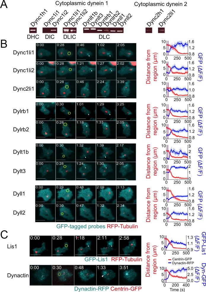
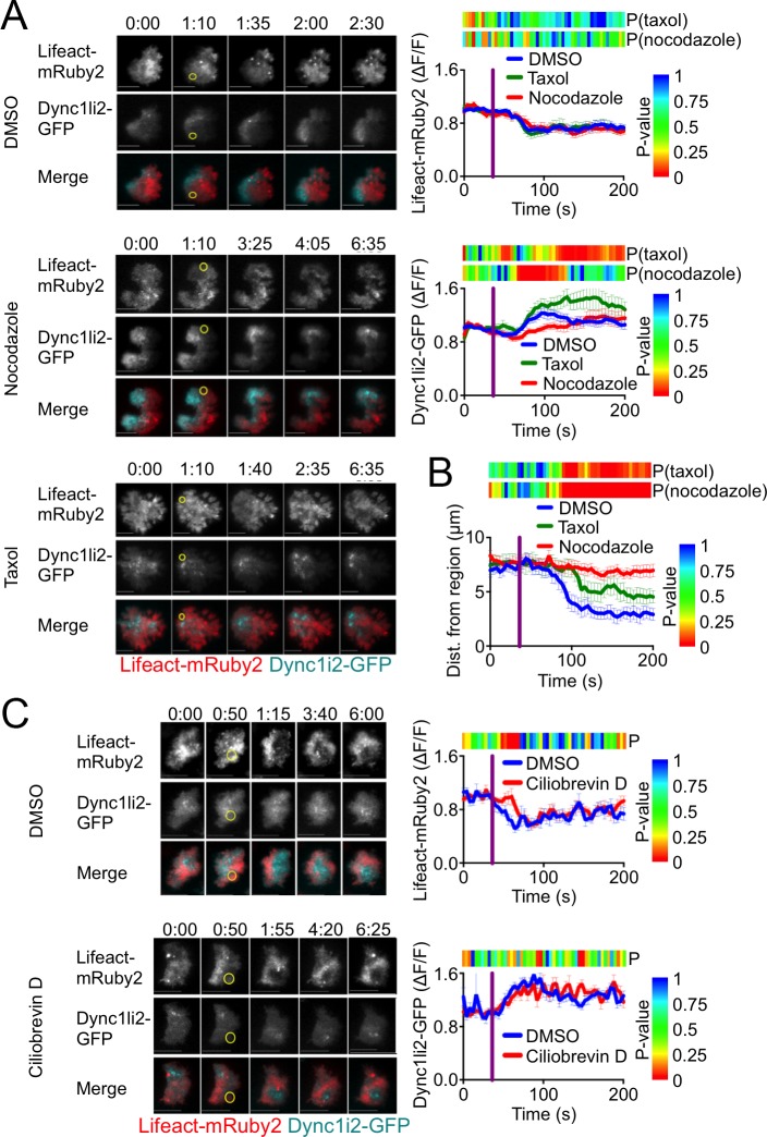
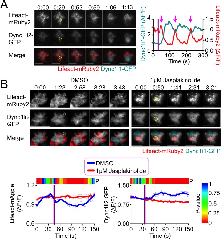
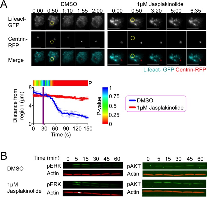
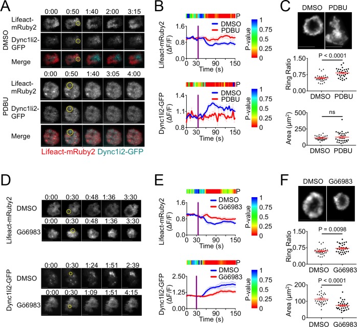
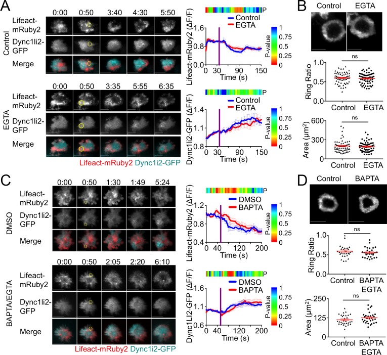
Immunological synapse (IS) formation between a T cell and an antigen-presenting cell is accompanied by the reorientation of the T cell centrosome toward the interface. This polarization response is thought to enhance the specificity of T cell effector function by enabling the directional secretion of cytokines and cytotoxic factors toward the antigen-presenting cell. Centrosome reorientation is controlled by polarized signaling through diacylglycerol (DAG) and protein kinase C (PKC). This drives the recruitment of the motor protein dynein to the IS, where it pulls on microtubules to reorient the centrosome. Here, we used T cell receptor photoactivation and imaging methodology to investigate the mechanisms controlling dynein accumulation at the synapse. Our results revealed a remarkable spatiotemporal correlation between dynein recruitment to the synaptic membrane and the depletion of cortical filamentous actin (F-actin) from the same region, suggesting that the two events were causally related. Consistent with this hypothesis, we found that pharmacological disruption of F-actin dynamics in T cells impaired both dynein accumulation and centrosome reorientation. DAG and PKC signaling were necessary for synaptic F-actin clearance and dynein accumulation, while calcium signaling and microtubules were dispensable for both responses. Taken together, these data provide mechanistic insight into the polarization of cytoskeletal regulators and highlight the close coordination between microtubule and F-actin architecture at the IS.

摘要

免疫突触(IS)形成于 T 细胞和抗原呈递细胞之间,伴随着 T 细胞中心体向界面的重新定向。这种极化反应被认为通过使细胞因子和细胞毒性因子朝向抗原呈递细胞的定向分泌来增强 T 细胞效应功能的特异性。中心体的重新定向由通过二酰基甘油(DAG)和蛋白激酶 C(PKC)的极化信号控制。这促使动力蛋白 dynein 向 IS 募集,在那里它拉动微管以重新定向中心体。在这里,我们使用 T 细胞受体光激活和成像方法来研究控制突触处 dynein 积累的机制。我们的结果揭示了 dynein 向突触膜募集与同一区域皮质丝状肌动蛋白(F-actin)耗竭之间的显著时空相关性,表明这两个事件存在因果关系。与该假设一致,我们发现 T 细胞中 F-actin 动力学的药理学破坏会损害 dynein 积累和中心体的重新定向。DAG 和 PKC 信号对于突触 F-actin 清除和 dynein 积累是必需的,而钙离子信号和微管对于这两种反应都是可有可无的。总之,这些数据提供了对细胞骨架调节剂极化的机制见解,并强调了微管和 F-actin 结构在 IS 处的紧密协调。

https://cdn.ncbi.nlm.nih.gov/pmc/blobs/038a/6608937/adc0d513f3c6/pone.0210377.g006.jpg
https://cdn.ncbi.nlm.nih.gov/pmc/blobs/038a/6608937/584741d48f9e/pone.0210377.g001.jpg
https://cdn.ncbi.nlm.nih.gov/pmc/blobs/038a/6608937/3f3ecb95a212/pone.0210377.g002.jpg
https://cdn.ncbi.nlm.nih.gov/pmc/blobs/038a/6608937/462b86da95bd/pone.0210377.g003.jpg
https://cdn.ncbi.nlm.nih.gov/pmc/blobs/038a/6608937/2c66dd1c1610/pone.0210377.g004.jpg
https://cdn.ncbi.nlm.nih.gov/pmc/blobs/038a/6608937/11626a1ab9c4/pone.0210377.g005.jpg
https://cdn.ncbi.nlm.nih.gov/pmc/blobs/038a/6608937/adc0d513f3c6/pone.0210377.g006.jpg

相似文献

1
Actin clearance promotes polarized dynein accumulation at the immunological synapse.肌动蛋白清除促进免疫突触极化的动力蛋白积累。
PLoS One. 2019 Jul 3;14(7):e0210377. doi: 10.1371/journal.pone.0210377. eCollection 2019.
2
Diacylglycerol promotes centrosome polarization in T cells via reciprocal localization of dynein and myosin II.二酰基甘油通过动力蛋白和肌球蛋白 II 的相互定位促进 T 细胞中心体极化。
Proc Natl Acad Sci U S A. 2013 Jul 16;110(29):11976-81. doi: 10.1073/pnas.1306180110. Epub 2013 Jul 1.
3
Kinesin-4 KIF21B limits microtubule growth to allow rapid centrosome polarization in T cells.动力蛋白-4 KIF21B 限制微管生长,以允许 T 细胞中快速的中心体极化。
Elife. 2020 Dec 21;9:e62876. doi: 10.7554/eLife.62876.
4
Dynein self-organizes while translocating the centrosome in T-cells.动力蛋白在 T 细胞中转运中心体时自我组织。
Mol Biol Cell. 2021 Apr 19;32(9):855-868. doi: 10.1091/mbc.E20-10-0668. Epub 2021 Mar 10.
5
A cascade of protein kinase C isozymes promotes cytoskeletal polarization in T cells.一连串的蛋白激酶 C 同工酶促进 T 细胞骨架极化。
Nat Immunol. 2011 May 22;12(7):647-54. doi: 10.1038/ni.2033.
6
Lymphocyte Polarization During Immune Synapse Assembly: Centrosomal Actin Joins the Game.免疫突触装配过程中的淋巴细胞极化:中心体肌动蛋白加入游戏。
Front Immunol. 2022 Feb 11;13:830835. doi: 10.3389/fimmu.2022.830835. eCollection 2022.
7
The Rap1-cofilin-1 pathway coordinates actin reorganization and MTOC polarization at the B cell immune synapse.Rap1-丝切蛋白-1信号通路在B细胞免疫突触处协调肌动蛋白重组和微管组织中心极化。
J Cell Sci. 2017 Mar 15;130(6):1094-1109. doi: 10.1242/jcs.191858. Epub 2017 Feb 6.
8
Analyzing Actin Dynamics at the Immunological Synapse.分析免疫突触处的肌动蛋白动力学。
Methods Mol Biol. 2017;1584:7-29. doi: 10.1007/978-1-4939-6881-7_2.
9
Centrosome repositioning in T cells is biphasic and driven by microtubule end-on capture-shrinkage.T 细胞中的中心体重定位是两相的,由微管末端捕获-收缩驱动。
J Cell Biol. 2013 Sep 2;202(5):779-92. doi: 10.1083/jcb.201301004. Epub 2013 Aug 26.
10
Diacylglycerol kinase ζ limits the polarized recruitment of diacylglycerol-enriched organelles to the immune synapse in T cells.二酰基甘油激酶ζ限制富含二酰基甘油的细胞器向T细胞免疫突触的极化募集。
Sci Signal. 2016 Dec 20;9(459):ra127. doi: 10.1126/scisignal.aaf7714.

引用本文的文献

1
Positioning centrioles and centrosomes.定位中心粒和中心体。
J Cell Biol. 2024 Apr 1;223(4). doi: 10.1083/jcb.202311140. Epub 2024 Mar 21.
2
Extracellular vesicles and microvilli in the immune synapse.免疫突触中的细胞外囊泡和微绒毛。
Front Immunol. 2024 Jan 3;14:1324557. doi: 10.3389/fimmu.2023.1324557. eCollection 2023.
3
Actin cytoskeleton remodeling at the cancer cell side of the immunological synapse: good, bad, or both?免疫突触中癌细胞侧的肌动蛋白细胞骨架重构:好、坏,还是皆有?

本文引用的文献

1
PIP5 Kinases Regulate Membrane Phosphoinositide and Actin Composition for Targeted Granule Secretion by Cytotoxic Lymphocytes.PIP5 激酶调节膜磷酯酰肌醇和肌动蛋白组成以靶向细胞毒性淋巴细胞的颗粒分泌。
Immunity. 2018 Sep 18;49(3):427-437.e4. doi: 10.1016/j.immuni.2018.08.017. Epub 2018 Sep 11.
2
Spatial and Temporal Control of T Cell Activation Using a Photoactivatable Agonist.使用光激活激动剂对T细胞活化进行时空控制。
J Vis Exp. 2018 Apr 25(134):56655. doi: 10.3791/56655.
3
Cutting Edge: Murine NK Cells Degranulate and Retain Cytotoxic Function without Store-Operated Calcium Entry.
Front Immunol. 2023 Oct 5;14:1276602. doi: 10.3389/fimmu.2023.1276602. eCollection 2023.
4
ALFA-PRF: a novel approach to detect murine perforin release from CTLs into the immune synapse.ALFA-PRF:一种新方法,用于检测 CTL 向免疫突触中释放的鼠穿孔素。
Front Immunol. 2022 Dec 22;13:931820. doi: 10.3389/fimmu.2022.931820. eCollection 2022.
5
T Lymphocyte and CAR-T Cell-Derived Extracellular Vesicles and Their Applications in Cancer Therapy.T 淋巴细胞和 CAR-T 细胞衍生的细胞外囊泡及其在癌症治疗中的应用。
Cells. 2022 Feb 24;11(5):790. doi: 10.3390/cells11050790.
6
Stochastic model of T cell repolarization during target elimination (II).目标消除过程中 T 细胞复极化的随机模型(二)。
Biophys J. 2022 Apr 5;121(7):1246-1265. doi: 10.1016/j.bpj.2022.02.029. Epub 2022 Feb 20.
7
Sorting Nexin 27 Enables MTOC and Secretory Machinery Translocation to the Immune Synapse.分选连接蛋白 27 使微管组织中心和分泌机器向免疫突触转运。
Front Immunol. 2022 Jan 12;12:814570. doi: 10.3389/fimmu.2021.814570. eCollection 2021.
8
Multifaceted roles of centrosomes in development, health, and disease.中心体在发育、健康和疾病中的多方面作用。
J Mol Cell Biol. 2021 Dec 6;13(9):611-621. doi: 10.1093/jmcb/mjab041.
9
F-Actin Dynamics in the Regulation of Endosomal Recycling and Immune Synapse Assembly.丝状肌动蛋白动力学在内体循环和免疫突触组装调控中的作用
Front Cell Dev Biol. 2021 Jun 24;9:670882. doi: 10.3389/fcell.2021.670882. eCollection 2021.
10
Dynein self-organizes while translocating the centrosome in T-cells.动力蛋白在 T 细胞中转运中心体时自我组织。
Mol Biol Cell. 2021 Apr 19;32(9):855-868. doi: 10.1091/mbc.E20-10-0668. Epub 2021 Mar 10.
前沿:小鼠自然杀伤细胞脱颗粒并保留细胞毒性功能,无需储存式钙内流。
J Immunol. 2017 Sep 15;199(6):1973-1978. doi: 10.4049/jimmunol.1700340. Epub 2017 Aug 9.
4
Cortical actin recovery at the immunological synapse leads to termination of lytic granule secretion in cytotoxic T lymphocytes.皮质肌动蛋白在免疫突触中的恢复导致细胞毒性 T 淋巴细胞中裂解颗粒分泌的终止。
Proc Natl Acad Sci U S A. 2017 Aug 8;114(32):E6585-E6594. doi: 10.1073/pnas.1710751114. Epub 2017 Jul 17.
5
Formin-generated actomyosin arcs propel T cell receptor microcluster movement at the immune synapse.formin生成的肌动球蛋白弧推动免疫突触处T细胞受体微簇的移动。
J Cell Biol. 2016 Nov 7;215(3):383-399. doi: 10.1083/jcb.201603080. Epub 2016 Oct 31.
6
Dynein Separately Partners with NDE1 and Dynactin To Orchestrate T Cell Focused Secretion.动力蛋白分别与NDE1和动力蛋白激活蛋白结合,以协调T细胞定向分泌。
J Immunol. 2016 Sep 15;197(6):2090-101. doi: 10.4049/jimmunol.1600180. Epub 2016 Aug 17.
7
Calcium influx through CRAC channels controls actin organization and dynamics at the immune synapse.通过钙释放激活钙通道(CRAC通道)的钙内流控制着免疫突触处肌动蛋白的组织和动力学。
Elife. 2016 Jul 21;5:e14850. doi: 10.7554/eLife.14850.
8
Mother Centriole Distal Appendages Mediate Centrosome Docking at the Immunological Synapse and Reveal Mechanistic Parallels with Ciliogenesis.母中心粒远端附属物介导免疫突触处的中心体对接,并揭示与纤毛发生的机制相似性。
Curr Biol. 2015 Dec 21;25(24):3239-44. doi: 10.1016/j.cub.2015.10.028. Epub 2015 Dec 5.
9
Comparative analysis of tools for live cell imaging of actin network architecture.肌动蛋白网络结构活细胞成像工具的比较分析
Bioarchitecture. 2014;4(6):189-202. doi: 10.1080/19490992.2014.1047714. Epub 2015 Aug 28.
10
Actin depletion initiates events leading to granule secretion at the immunological synapse.肌动蛋白的消耗引发了导致免疫突触处颗粒分泌的一系列事件。
Immunity. 2015 May 19;42(5):864-76. doi: 10.1016/j.immuni.2015.04.013.