Takatsuna Masafumi, Takeuchi Manabu, Usuda Hiroyuki, Terai Shuji
Department of Gastroenterology, Nagaoka Red Cross Hospital, Nagaoka, Japan.
Department of Pathology, Nagaoka Red Cross Hospital, Nagaoka, Japan.
Endosc Int Open. 2019 Jul;7(7):E893-E895. doi: 10.1055/a-0918-5804. Epub 2019 Jul 3.
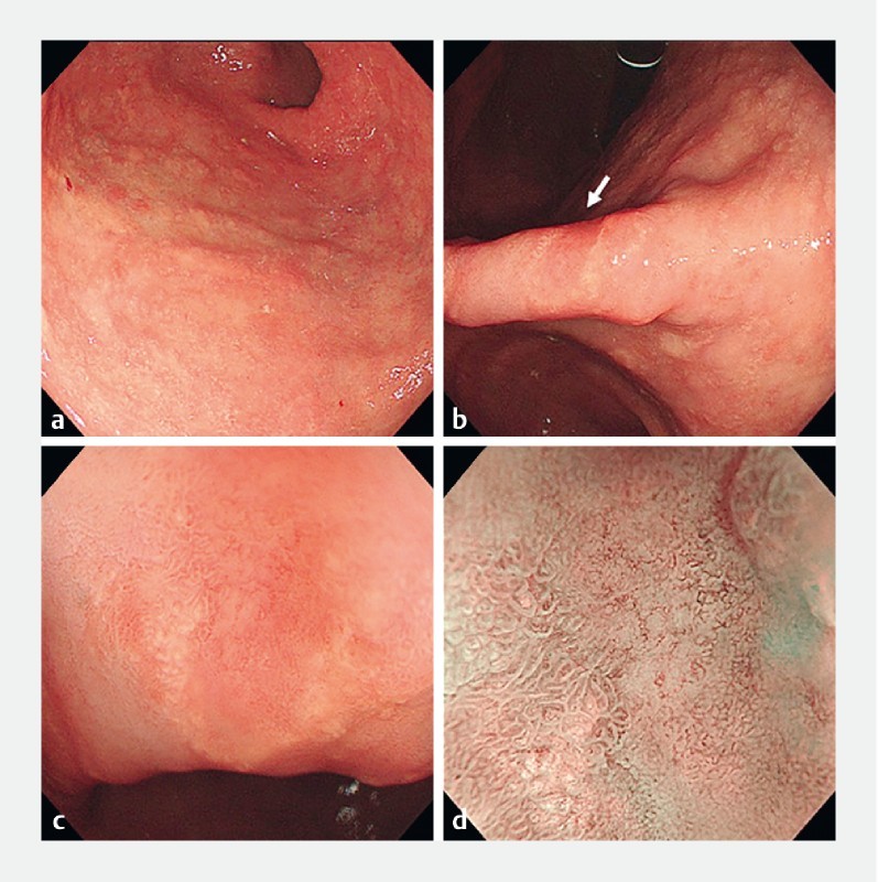
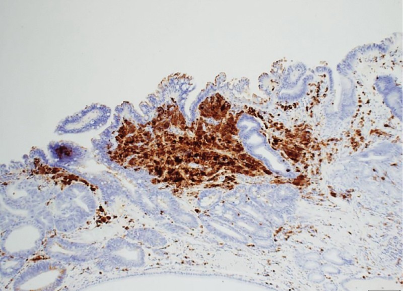
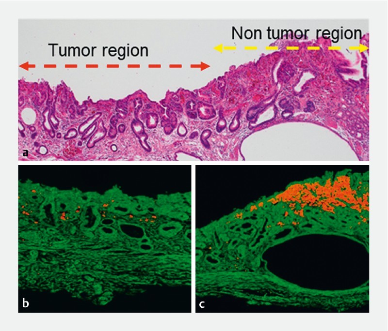
A 78-year-old man with infection had been undergoing hemodialysis for chronic renal failure and receiving lanthanum carbonate preparations for 3 years. Endoscopy revealed fine white granular discoloration throughout the stomach, a moderately reddish depression on the lesser curvature of the gastric angle, and white granular discoloration in the surrounding area. A magnified image using narrow-band imaging showed that the depressed part had irregular vascular and pit structures. We established a diagnosis of intramucosal gastric cancer and performed endoscopic submucosal dissection. Histopathological examination revealed a well-differentiated adenocarcinoma that was confined to the mucosa of the depressed area. Moreover, using an electron probe microanalyzer-equipped electron microscope, we found that the degree of lanthanum deposition was lower in the tumor region than in the non-tumor region. Thus, the current case can help in understanding the relationship between lanthanum deposition and early-stage gastric cancer. Because gastric cancers can occur in lanthanum deposit-containing mucosa, esophagogastroduodenoscopy should be used carefully after understanding the characteristics of early- stage gastric cancer in such cases.
一名78岁的感染患者因慢性肾衰竭一直在接受血液透析,并服用碳酸镧制剂3年。内镜检查显示整个胃黏膜呈细小白色颗粒状变色,胃角小弯处有中度发红的凹陷,周围区域有白色颗粒状变色。使用窄带成像的放大图像显示,凹陷部分有不规则的血管和凹陷结构。我们诊断为黏膜内胃癌,并进行了内镜黏膜下剥离术。组织病理学检查显示为高分化腺癌,局限于凹陷区域的黏膜。此外,使用配备电子探针微分析仪的电子显微镜,我们发现肿瘤区域的镧沉积程度低于非肿瘤区域。因此,本病例有助于理解镧沉积与早期胃癌之间的关系。由于胃癌可发生在含镧沉积的黏膜中,因此在了解此类病例中早期胃癌的特征后,应谨慎使用食管胃十二指肠镜检查。