Zhu Yong-Jian, Feng Bing, Wang Shuang, Wang Li-Ming, Wu Jiang-Fen, Ma Xiao-Hong, Zhao Xin-Ming
Department of Imaging Diagnosis, National Cancer Center/National Clinical Research Center for Cancer/Cancer Hospital, Chinese Academy of Medical Sciences and Peking Union Medical College, Beijing 100021, P.R. China.
Department of Hepatobiliary Surgery, National Cancer Center/National Clinical Research Center for Cancer/Cancer Hospital, Chinese Academy of Medical Sciences and Peking Union Medical College, Beijing 100021, P.R. China.
Oncol Lett. 2019 Jul;18(1):720-732. doi: 10.3892/ol.2019.10378. Epub 2019 May 20.
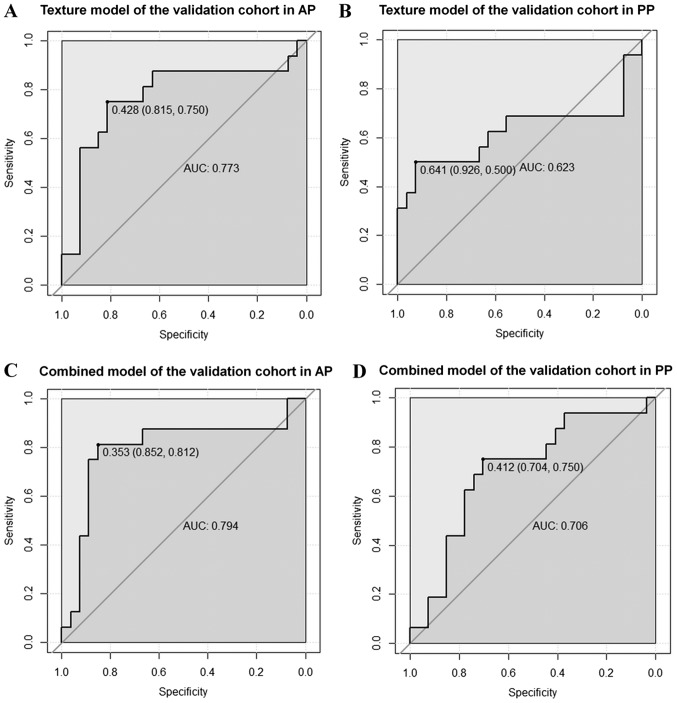
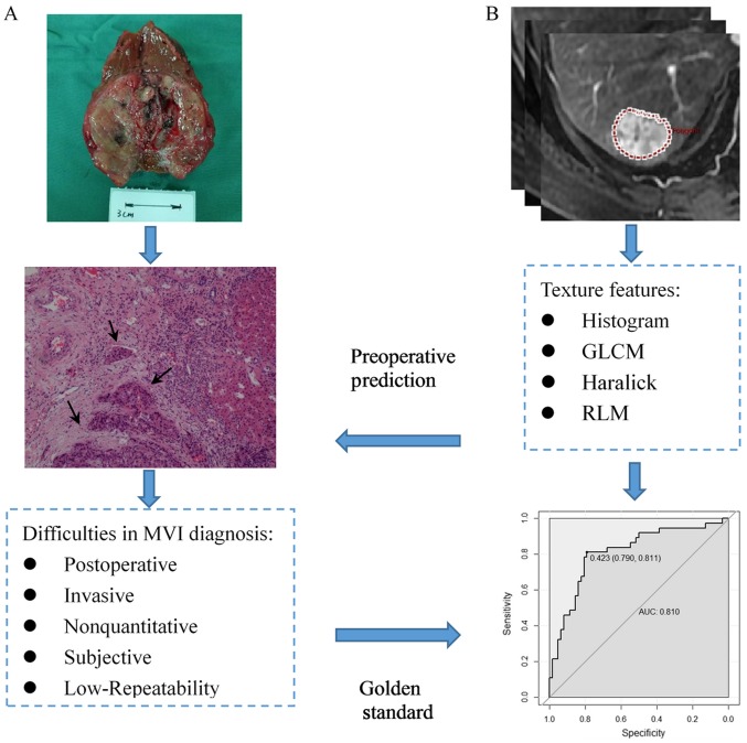
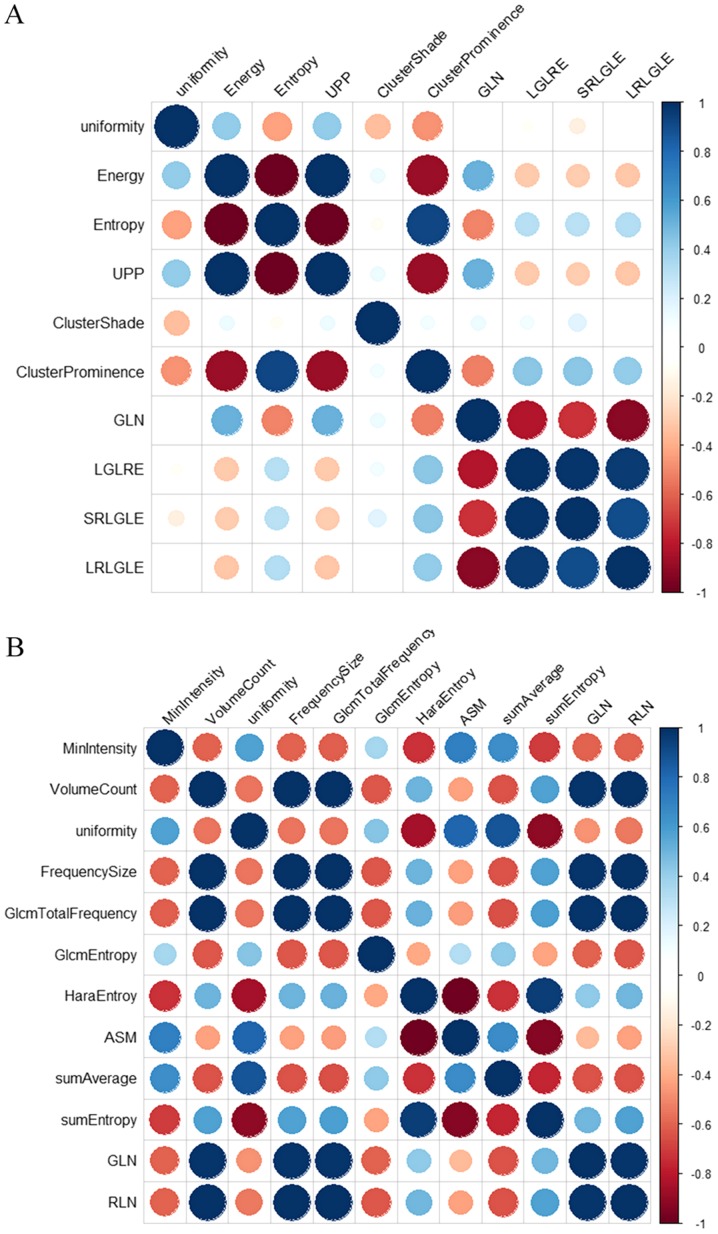
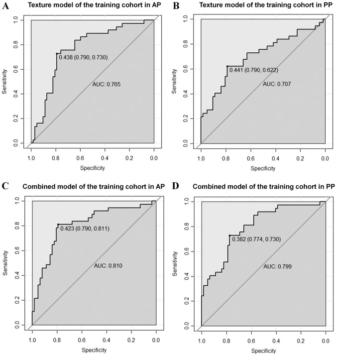
The purpose of the present study was to investigate the value of contrast-enhanced magnetic resonance imaging (CE-MRI) texture analysis for preoperatively predicting microvascular invasion (MVI) in hepatocellular carcinoma (HCC). Accordingly, a retrospective study of 142 patients with pathologically confirmed HCC was performed. The patients were divided into two cohorts: The training cohort (n=99) and the validation cohort (n=43), including the MVI-positive group (n=53) and MVI-negative group (n=89). On the basis of three-dimensional texture analysis, 58 features were extracted from the preoperative CE-MR images of arterial-phase (AP) and portal-venous-phase (PP). The t-test or Kruskal-Wallis test, univariate logistic regression analysis and Pearson correlation were applied for feature reduction. Clinical-radiological features were also analyzed. Multivariate logistic regression analysis was used to build the texture model and combined model with clinical-radiological features. The MVI-predictive performance of the models was evaluated using receiver operating characteristic (ROC) analysis and presented using nomogram. Among the clinical features, a significant difference was found in maximum tumor diameter (P=0.002), tumor differentiation (P=0.026) and α-fetoprotein level (P=0.025) between the two groups in the training cohort. Four MR texture features in AP and five in PP images were identified through feature reduction. On ROC analysis, the AP texture model showed better diagnostic performance than did the PP model in the validation cohort, with an area under the curve (AUC) of 0.773 vs. 0.623, sensitivity of 0.750 vs. 0.500, and specificity of 0.815 vs. 0.926. Together with the clinical features, the combined model of AP improved the AUC, sensitivity and specificity to 0.810, 0.811 and 0.790, respectively, which was demonstrated in nomogram. To conclude, model-based texture analysis of CE-MRI could predict MVI in HCC preoperatively and noninvasively, and the AP image shows better predictive efficiency than PP image. The combined model of AP with clinical-radiological features could improve MVI prediction ability.
本研究的目的是探讨对比增强磁共振成像(CE-MRI)纹理分析对术前预测肝细胞癌(HCC)微血管侵犯(MVI)的价值。因此,对142例经病理证实的HCC患者进行了回顾性研究。患者被分为两个队列:训练队列(n=99)和验证队列(n=43),包括MVI阳性组(n=53)和MVI阴性组(n=89)。基于三维纹理分析,从动脉期(AP)和门静脉期(PP)的术前CE-MR图像中提取了58个特征。采用t检验或Kruskal-Wallis检验、单因素逻辑回归分析和Pearson相关性分析进行特征降维。还分析了临床放射学特征。采用多因素逻辑回归分析建立纹理模型以及结合临床放射学特征的联合模型。使用受试者工作特征(ROC)分析评估模型对MVI的预测性能,并通过列线图展示。在临床特征方面,训练队列中两组之间的最大肿瘤直径(P=0.002)、肿瘤分化程度(P=0.026)和甲胎蛋白水平(P=0.025)存在显著差异。通过特征降维,在AP图像中识别出4个MR纹理特征,在PP图像中识别出5个。在ROC分析中,验证队列中AP纹理模型的诊断性能优于PP模型,曲线下面积(AUC)分别为0.773和0.623,灵敏度分别为0.750和0.500,特异性分别为0.815和0.926。与临床特征相结合,AP联合模型将AUC、灵敏度和特异性分别提高到0.810、0.811和0.790,列线图显示了这一结果。总之,基于模型的CE-MRI纹理分析可以术前无创预测HCC中的MVI,并且AP图像的预测效率优于PP图像。AP与临床放射学特征的联合模型可以提高MVI预测能力。