Department of Radiology, St. Luke's International Hospital.
Department of Radiology, Keio University School of Medicine.
Magn Reson Med Sci. 2020 Aug 3;19(3):282-285. doi: 10.2463/mrms.cr.2019-0051. Epub 2019 Jul 11.
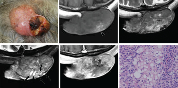
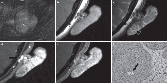
Few studies had been published regarding imaging findings of skin adnexal tumors. We experienced two giant cases of them with a characteristic mushroom-like growth pattern. MRI showed a circumscribed mushroom-like shaped mass extruding from the subcutaneous tissue with microcystic lesions. Although differentiation between benignancy and malignancy may be difficult by radiological examinations, MRI may be helpful to identify its origin and differentiate soft tissue tumors with skin adnexal tumors in having these imaging findings.
关于皮肤附属器肿瘤的影像学表现,目前仅有少数研究报道。我们遇到了两例具有特征性蘑菇样生长模式的巨大病例。MRI 显示一个边界清楚的蘑菇状肿块从皮下组织向外突出,伴有微囊状病变。虽然影像学检查可能难以区分良恶性,但 MRI 可能有助于确定其来源,并在具有这些影像学表现的软组织肿瘤中区分皮肤附属器肿瘤。