Department of Psychiatry, Psychotherapy and Psychosomatics, Faculty of Medicine, RWTH Aachen, Aachen, Germany.
Institute of Neuroscience and Medicine: JARA-Institute Brain Structure Function Relationship (INM 10), Research Center Jülich, Jülich, Germany.
Hum Brain Mapp. 2019 Oct 15;40(15):4470-4486. doi: 10.1002/hbm.24715. Epub 2019 Jul 13.
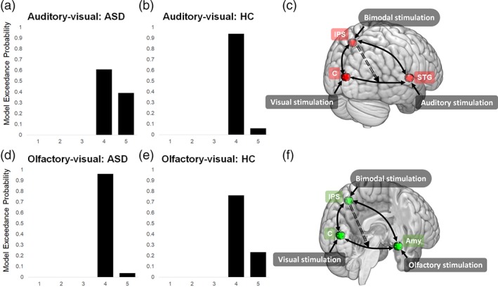
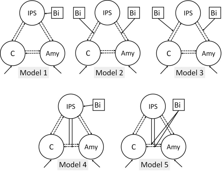
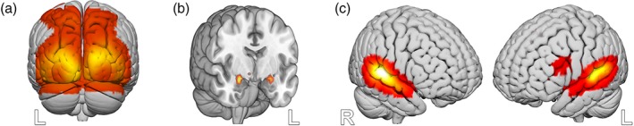
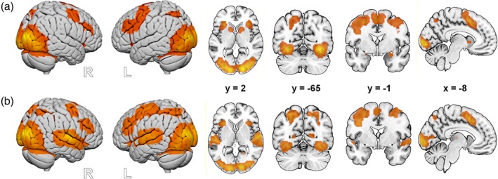
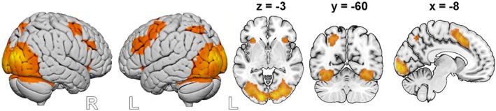
The human capacity to integrate sensory signals has been investigated with respect to different sensory modalities. A common denominator of the neural network underlying the integration of sensory clues has yet to be identified. Additionally, brain imaging data from patients with autism spectrum disorder (ASD) do not cover disparities in neuronal sensory processing. In this fMRI study, we compared the underlying neural networks of both olfactory-visual and auditory-visual integration in patients with ASD and a group of matched healthy participants. The aim was to disentangle sensory-specific networks so as to derive a potential (amodal) common source of multisensory integration (MSI) and to investigate differences in brain networks with sensory processing in individuals with ASD. In both groups, similar neural networks were found to be involved in the olfactory-visual and auditory-visual integration processes, including the primary visual cortex, the inferior parietal sulcus (IPS), and the medial and inferior frontal cortices. Amygdala activation was observed specifically during olfactory-visual integration, with superior temporal activation having been seen during auditory-visual integration. A dynamic causal modeling analysis revealed a nonlinear top-down IPS modulation of the connection between the respective primary sensory regions in both experimental conditions and in both groups. Thus, we demonstrate that MSI has shared neural sources across olfactory-visual and audio-visual stimulation in patients and controls. The enhanced recruitment of the IPS to modulate changes between areas is relevant to sensory perception. Our results also indicate that, with respect to MSI processing, adults with ASD do not significantly differ from their healthy counterparts.
人类整合感觉信号的能力已经在不同的感觉模式方面进行了研究。尚未确定整合感觉线索的神经网络的共同特征。此外,自闭症谱系障碍(ASD)患者的脑成像数据并未涵盖神经元感觉处理的差异。在这项 fMRI 研究中,我们比较了 ASD 患者和一组匹配的健康参与者的嗅觉-视觉和听觉-视觉整合的基础神经网络。目的是分离出感觉特异性网络,以便得出多感觉整合(MSI)的潜在(非模态)共同来源,并研究 ASD 个体中具有感觉处理的脑网络的差异。在两组中,都发现相似的神经网络参与了嗅觉-视觉和听觉-视觉整合过程,包括初级视觉皮层、下顶叶沟(IPS)、内侧和下额皮质。在嗅觉-视觉整合过程中观察到杏仁核激活,在听觉-视觉整合过程中观察到颞上回激活。动态因果建模分析显示,在两种实验条件和两组中,顶叶 IPS 对各自初级感觉区域之间的连接进行非线性的自上而下的调制。因此,我们证明了 MSI 在患者和对照组的嗅觉-视觉和听觉-视觉刺激中具有共同的神经来源。IPS 的增强募集来调节区域之间的变化与感觉感知有关。我们的结果还表明,在 MSI 处理方面,ASD 成人与他们的健康对照组没有显著差异。