Institute of Immunology, Friedrich-Loeffler-Institut, Greifswald-Insel Riems, Germany.
Department of Experimental Animal Facilities and Biorisk Management, Friedrich-Loeffler-Institut, Greifswald-Insel Riems, Germany.
Front Immunol. 2019 Jun 18;10:1380. doi: 10.3389/fimmu.2019.01380. eCollection 2019.
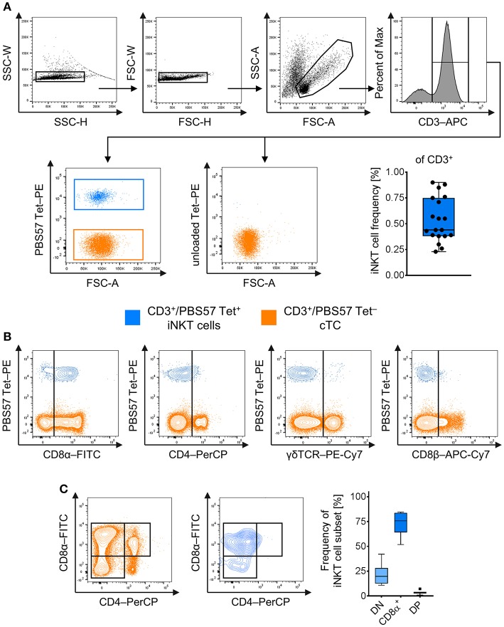
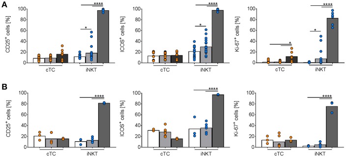
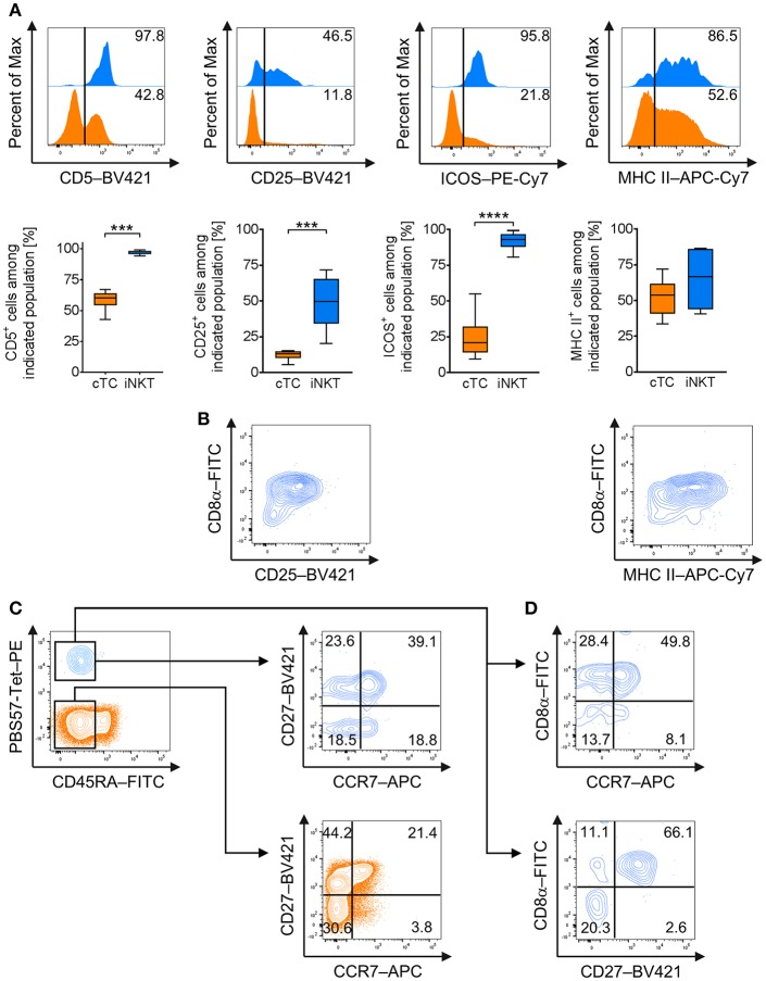
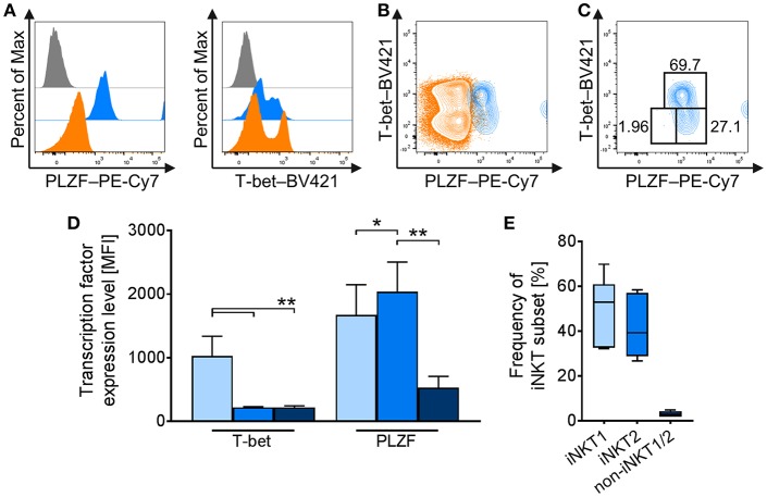
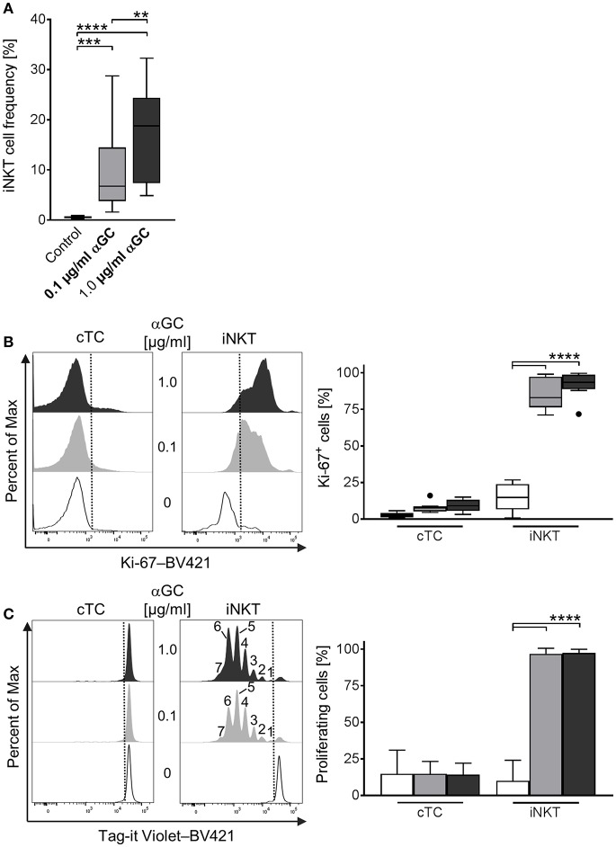
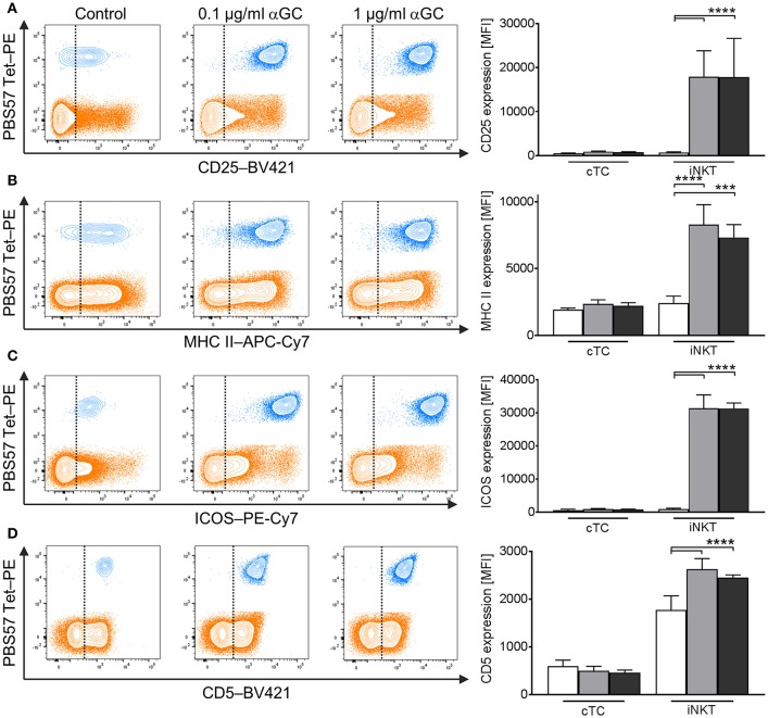
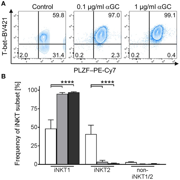
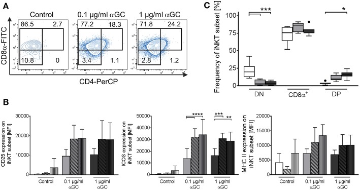
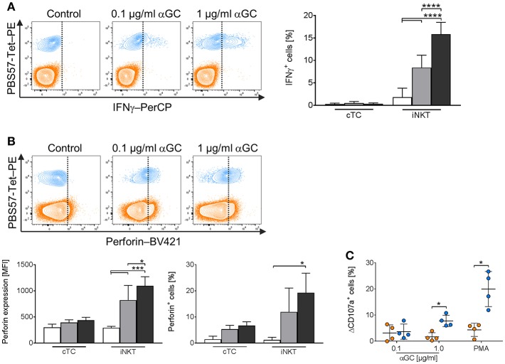
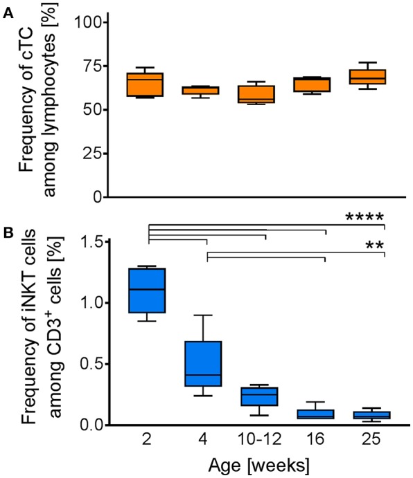
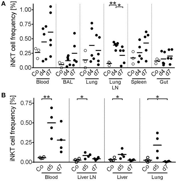
Pigs are important livestock and comprehensive understanding of their immune responses in infections is critical to improve vaccines and therapies. Moreover, similarities between human and swine physiology suggest that pigs are a superior animal model for immunological studies. However, paucity of experimental tools for a systematic analysis of the immune responses in pigs represent a major disadvantage. To evaluate the pig as a biomedical model and additionally expand the knowledge of rare immune cell populations in swine, we established a multicolor flow cytometry analysis platform of surface marker expression and cellular responses for porcine invariant Natural Killer T cells (iNKT). In humans, iNKT cells are among the first line defenders in various tissues, respond to CD1d-restricted antigens and become rapidly activated. Naïve porcine iNKT cells were CD3/CD4/CD8 or CD3/CD4/CD8 and displayed an effector- or memory-like phenotype (CD25/ICOS/CD5/CD45RA/CCR7 /CD27). Based on their expression of the transcription factors T bet and the iNKT cell-specific promyelocytic leukemia zinc finger protein (PLZF), porcine iNKT cells were differentiated into functional subsets. Analogous to human iNKT cells, stimulation of porcine leukocytes with the CD1d ligand α-galactosylceramide resulted in rapid iNKT cell proliferation, evidenced by an increase in frequency and Ki-67 expression. Moreover, this approach revealed CD25, CD5, ICOS, and the major histocompatibility complex class II (MHC II) as activation markers on porcine iNKT cells. Activated iNKT cells also expressed interferon-γ, upregulated perforin expression, and displayed degranulation. In steady state, iNKT cell frequency was highest in newborn piglets and decreased with age. Upon infection with two viruses of high relevance to swine and humans, iNKT cells expanded. Animals infected with African swine fever virus displayed an increase of iNKT cell frequency in peripheral blood, regional lymph nodes, and lungs. During Influenza A virus infection, iNKT cell percentage increased in blood, lung lymph nodes, and broncho-alveolar lavage. Our in-depth characterization of porcine iNKT cells contributes to a better understanding of porcine immune responses, thereby facilitating the design of innovative interventions against infectious diseases. Moreover, we provide new evidence that endorses the suitability of the pig as a biomedical model for iNKT cell research.
猪是重要的家畜,全面了解其感染时的免疫反应对于改进疫苗和治疗方法至关重要。此外,人类和猪的生理学相似性表明,猪是免疫研究的优越动物模型。然而,用于系统分析猪免疫反应的实验工具的缺乏代表了一个主要的缺点。为了评估猪作为生物医学模型,并进一步扩展猪中稀有免疫细胞群体的知识,我们建立了用于猪不变自然杀伤 T 细胞(iNKT)的表面标记表达和细胞反应的多色流式细胞术分析平台。在人类中,iNKT 细胞是各种组织中第一线防御者之一,对 CD1d 限制的抗原作出反应,并迅速被激活。幼稚的猪 iNKT 细胞是 CD3/CD4/CD8 或 CD3/CD4/CD8,表现出效应器或记忆样表型(CD25/ICOS/CD5/CD45RA/CCR7/CD27)。基于其转录因子 T bet 和 iNKT 细胞特异性早幼粒细胞白血病锌指蛋白(PLZF)的表达,猪 iNKT 细胞被分化为功能性亚群。类似于人类 iNKT 细胞,用 CD1d 配体 α-半乳糖基神经酰胺刺激猪白细胞导致 iNKT 细胞快速增殖,这表现为频率增加和 Ki-67 表达增加。此外,这种方法揭示了 CD25、CD5、ICOS 和主要组织相容性复合体 II(MHC II)作为猪 iNKT 细胞的激活标记物。活化的 iNKT 细胞还表达干扰素-γ,上调穿孔素表达,并显示脱颗粒。在稳定状态下,iNKT 细胞频率在新生仔猪中最高,并随年龄增长而降低。感染两种对猪和人类具有高度相关性的病毒后,iNKT 细胞扩增。感染非洲猪瘟病毒的动物在外周血、局部淋巴结和肺中显示 iNKT 细胞频率增加。在流感病毒 A 感染期间,iNKT 细胞百分比在血液、肺淋巴结和支气管肺泡灌洗中增加。我们对猪 iNKT 细胞的深入表征有助于更好地理解猪的免疫反应,从而促进针对传染病的创新干预措施的设计。此外,我们提供了新的证据,支持猪作为 iNKT 细胞研究的生物医学模型的适宜性。