Zhang Jun, Gao Yali
1Department of Obstetrics and Gynecology, The Second Clinical Medical College (Shenzhen People's Hospital), Jinan University, Shenzhen, 518020 People's Republic of China.
2Department of Ophthalmology, The Second Clinical Medical College (Shenzhen People's Hospital), Jinan University, Shenzhen, 518020 People's Republic of China.
Cancer Cell Int. 2019 Jul 8;19:175. doi: 10.1186/s12935-019-0893-z. eCollection 2019.
Maternally expressed 3 (MEG3) plays an important role in cervical cancer development, but its exact role remains unclear. Here, we explored the specific regulatory mechanism of MEG3 and its downstream proteins in cervical cancer cells.
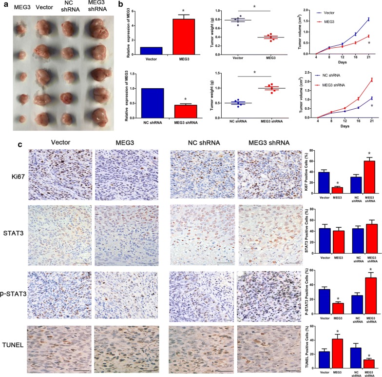
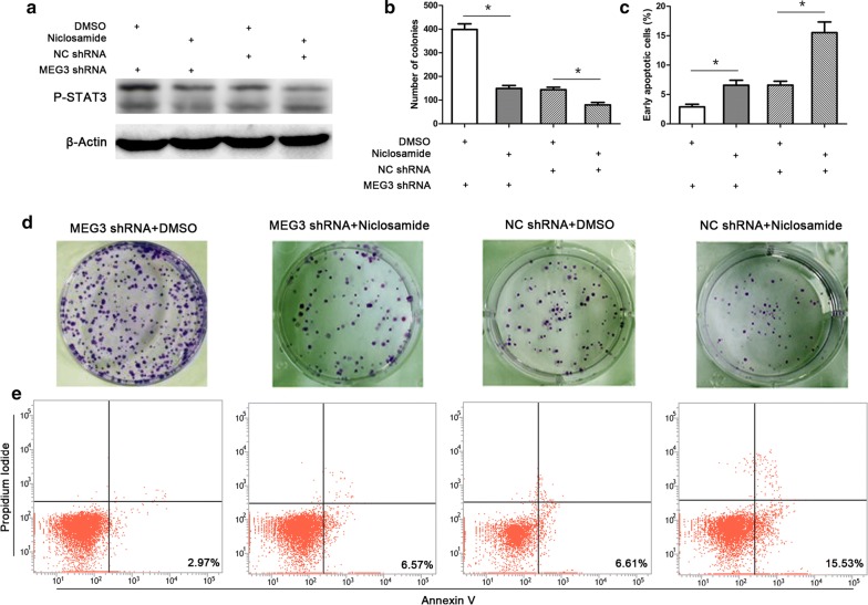
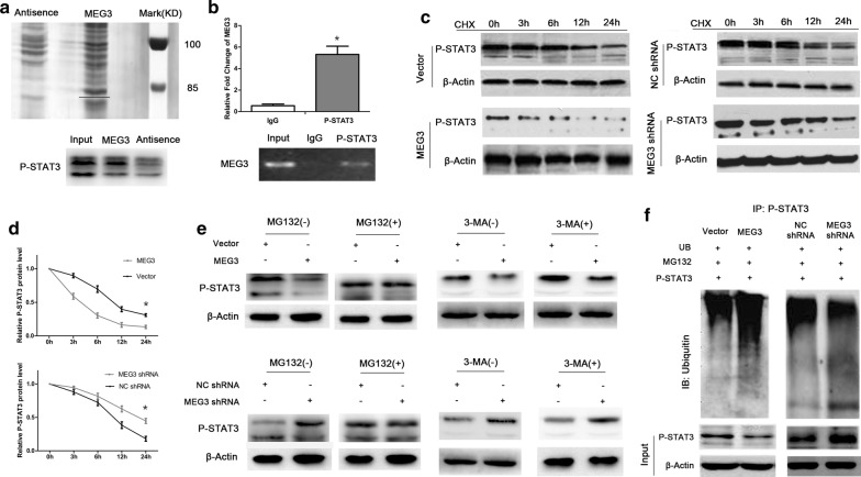
The effect of MEG3 on tumor formation ability of cervical cancer cells was determined in nude mice. The direct binding of MEG3 to phosphorylated signal transducer and activator of transcription 3 (P-STAT3) was detected by RNA pull-down and RNA-binding protein immunoprecipitation (RIP) assays. Cycloheximide (CHX)-chase and ubiquitination assays were performed to determine the regulatory effect of MEG3 on P-STAT3 ubiquitination. Clone formation assay and flow cytometry were used to evaluate the effect of the MEG3-STAT3 regulatory axis on cell proliferation and apoptosis.
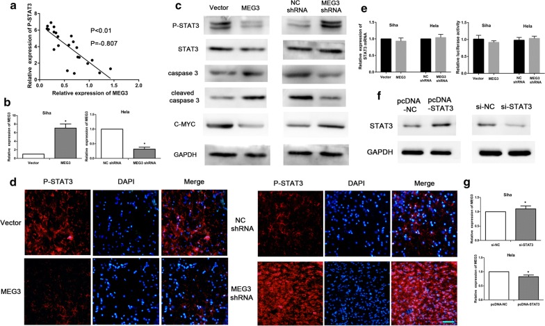
In vivo tumor formation experiments showed that MEG3 inhibited the tumor formation ability of cervical cancer cells. RNA pull-down and RIP assays demonstrated that MEG3 bound directly to P-STAT3 protein. CHX-chase and ubiquitination assay results showed that MEG3 promoted P-STAT3 degradation via ubiquitination. Clone formation assay and flow cytometry analysis results revealed that the inhibitory effect of MEG3 on P-STAT3 promoted apoptosis and inhibited proliferation of cervical cancer cells.
MEG3 binds to P-STAT3 in cervical cancer cells, resulting in P-STAT3 ubiquitination and degradation and apoptosis and inhibition of proliferation of tumor cells. The in-depth elaboration of the MEG3-STAT3 regulatory axis in cervical cancer may clarify the mechanism of action of MEG3 and provide new ideas for cervical cancer treatment.
母源表达基因3(MEG3)在宫颈癌发展过程中发挥重要作用,但其确切作用仍不清楚。在此,我们探讨了MEG3及其下游蛋白在宫颈癌细胞中的具体调控机制。
在裸鼠中确定MEG3对宫颈癌细胞肿瘤形成能力的影响。通过RNA下拉和RNA结合蛋白免疫沉淀(RIP)试验检测MEG3与磷酸化信号转导和转录激活因子3(P-STAT3)的直接结合。进行环己酰亚胺(CHX)追踪和泛素化试验以确定MEG3对P-STAT3泛素化的调控作用。采用克隆形成试验和流式细胞术评估MEG3-STAT3调控轴对细胞增殖和凋亡的影响。
体内肿瘤形成实验表明,MEG3抑制宫颈癌细胞的肿瘤形成能力。RNA下拉和RIP试验表明MEG3直接与P-STAT3蛋白结合。CHX追踪和泛素化试验结果表明,MEG3通过泛素化促进P-STAT3降解。克隆形成试验和流式细胞术分析结果显示,MEG3对P-STAT3的抑制作用促进了宫颈癌细胞的凋亡并抑制了其增殖。
MEG3在宫颈癌细胞中与P-STAT3结合,导致P-STAT3泛素化和降解,以及肿瘤细胞凋亡和增殖抑制。深入阐述宫颈癌中MEG3-STAT3调控轴可能阐明MEG3的作用机制,并为宫颈癌治疗提供新思路。