Park Jae Bum, Lee Song Am, Lee Woo Surng, Kim Yo Han, Song Inyoung, Lee Jeong Geun, Hwang Jae Joon
Department of Thoracic and Cardiovascular Surgery, Konkuk University Medical Center, Seoul, South Korea.
Department of Radiology, Konkuk University Medical Center, Seoul, South Korea.
Ann Thorac Med. 2019 Jul-Sep;14(3):205-212. doi: 10.4103/atm.ATM_287_18.
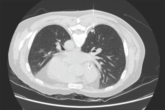
Confirming the histologic diagnosis of small pulmonary nodules or Ground-glass opacity nodules (GGNs) of unknown origin is difficult. These nodules are not always appropriate for percutaneous transthoracic needle biopsy. Preoperative localization of pulmonary lesions provides more precise target points to ensure complete surgical excision. The goal of the present study was to evaluate the validity and effectiveness of computed tomography-guided preoperative hook wire localization with our technique for video-assisted thoracoscopic surgery (VATS).
We retrospectively investigated 113 patients who had undergone preoperative hook wire localization before VATS resection for newly present or growing pulmonary nodular lesions between May 2007 and December 2016. Procedural and perioperative outcomes were assessed to evaluate the safety and efficacy of preoperative localization technique.
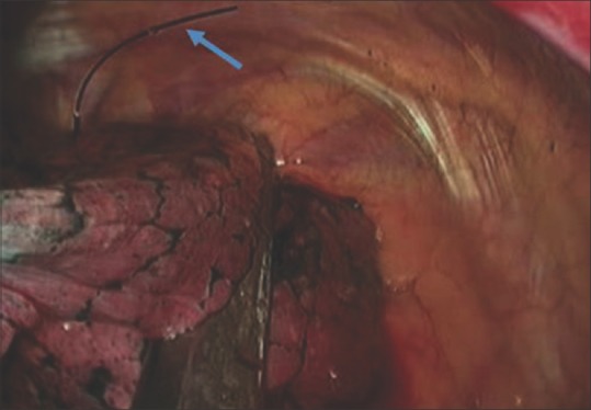
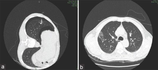
A total of 113 pulmonary nodules were localized and successfully resected in all 113 patients. The mean diameter of nodules was 10.8 ± 6.1 mm (range, 3-28). The mean distance from the pleural surface was 20.2 ± 12.4 mm (range, 5-55). The mean procedure time of localization was 23.7 ± 6.3 min. Asymptomatic minimal pneumothorax and mild parenchymal hemorrhage occurred in 26 (23.0%) and 8 (7.1%) patients, respectively. There were 32 (28.3%) deep lung nodules, in which the distance to pleural surface was more than 25 mm. Wire dislodgement occurred in 4 (3.5%) patients. Complete resection of all lung lesions was achieved, and definite histological diagnosis was obtained in all patients. Pathologic examination revealed 42 (37.2%) primary lung cancers, 2 (1.8%) lymphomas, 53 (46.9%) metastases, 16 (14.1%) benign lesions.
Preoperative percutaneous hook wire localization is a dependable and useful technique to facilitate positioning small and deep pulmonary nodules for thoracoscopic complete excision and accurate diagnosis.
对来源不明的小肺结节或磨玻璃密度结节(GGNs)进行组织学诊断颇具难度。这些结节并不总是适合经皮经胸针吸活检。肺部病变的术前定位可提供更精确的靶点,以确保手术完整切除。本研究的目的是评估采用我们的技术进行计算机断层扫描引导下的术前钩丝定位在电视辅助胸腔镜手术(VATS)中的有效性和安全性。
我们回顾性研究了2007年5月至2016年12月期间因新出现或增大的肺结节性病变而在VATS切除术前接受术前钩丝定位的113例患者。评估手术过程及围手术期结果,以评价术前定位技术的安全性和有效性。
113例患者的113个肺结节均成功定位并切除。结节的平均直径为10.8±6.1mm(范围3 - 28mm)。距胸膜表面的平均距离为20.2±12.4mm(范围5 - 55mm)。定位的平均操作时间为23.7±6.3分钟。分别有26例(23.0%)和8例(7.1%)患者出现无症状的少量气胸和轻度实质内出血。有32个(28.3%)深部肺结节,其距胸膜表面的距离超过25mm。4例(3.5%)患者出现钢丝移位。所有肺部病变均实现完整切除,所有患者均获得明确的组织学诊断。病理检查显示42例(37.2%)原发性肺癌、2例(1.8%)淋巴瘤、53例(46.9%)转移瘤、16例(14.1%)良性病变。
术前经皮钩丝定位是一种可靠且有用的技术,有助于对小的深部肺结节进行定位,以便胸腔镜完整切除并准确诊断。