From the Queens Medical Research Institute, BHF Centre for Cardiovascular Sciences, University of Edinburgh, United Kingdom (A.D.M., M.D.B., V.M., K.P., J.H., J.P.S., J.I., J.K., A.S.T., N.L.M., D.E.N., A.C., J.C.S., J.R., A.H.B.).
Institute of Cardiovascular and Medical Sciences, BHF Cardiovascular Research Centre, University of Glasgow, United Kingdom (A.C.B.).
Circ Res. 2019 Aug 16;125(5):535-551. doi: 10.1161/CIRCRESAHA.119.314876. Epub 2019 Jul 24.
In response to blood vessel wall injury, aberrant proliferation of vascular smooth muscle cells (SMCs) causes pathological remodeling. However, the controlling mechanisms are not completely understood.
We recently showed that the human long noncoding RNA, SMILR, promotes vascular SMCs proliferation by a hitherto unknown mechanism. Here, we assess the therapeutic potential of SMILR inhibition and detail the molecular mechanism of action.
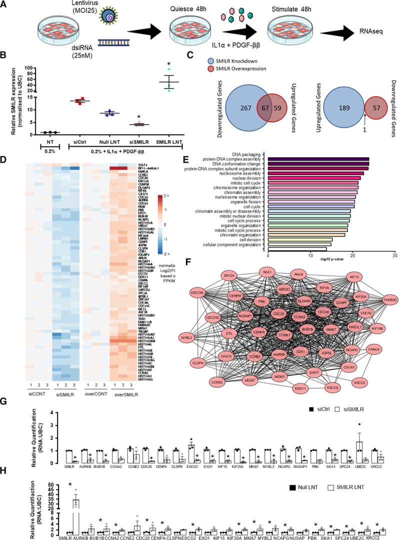
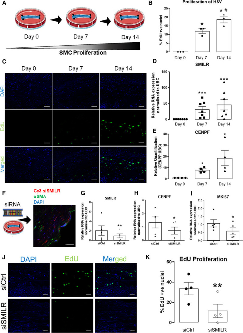
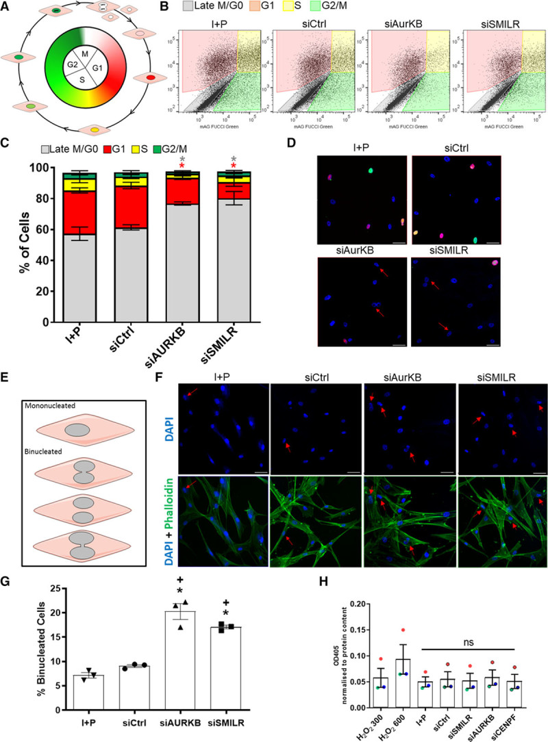
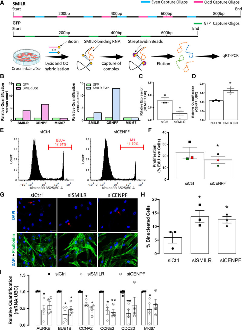
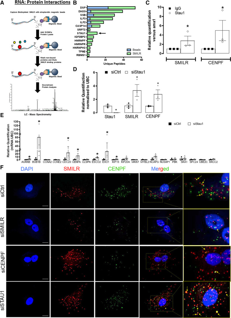
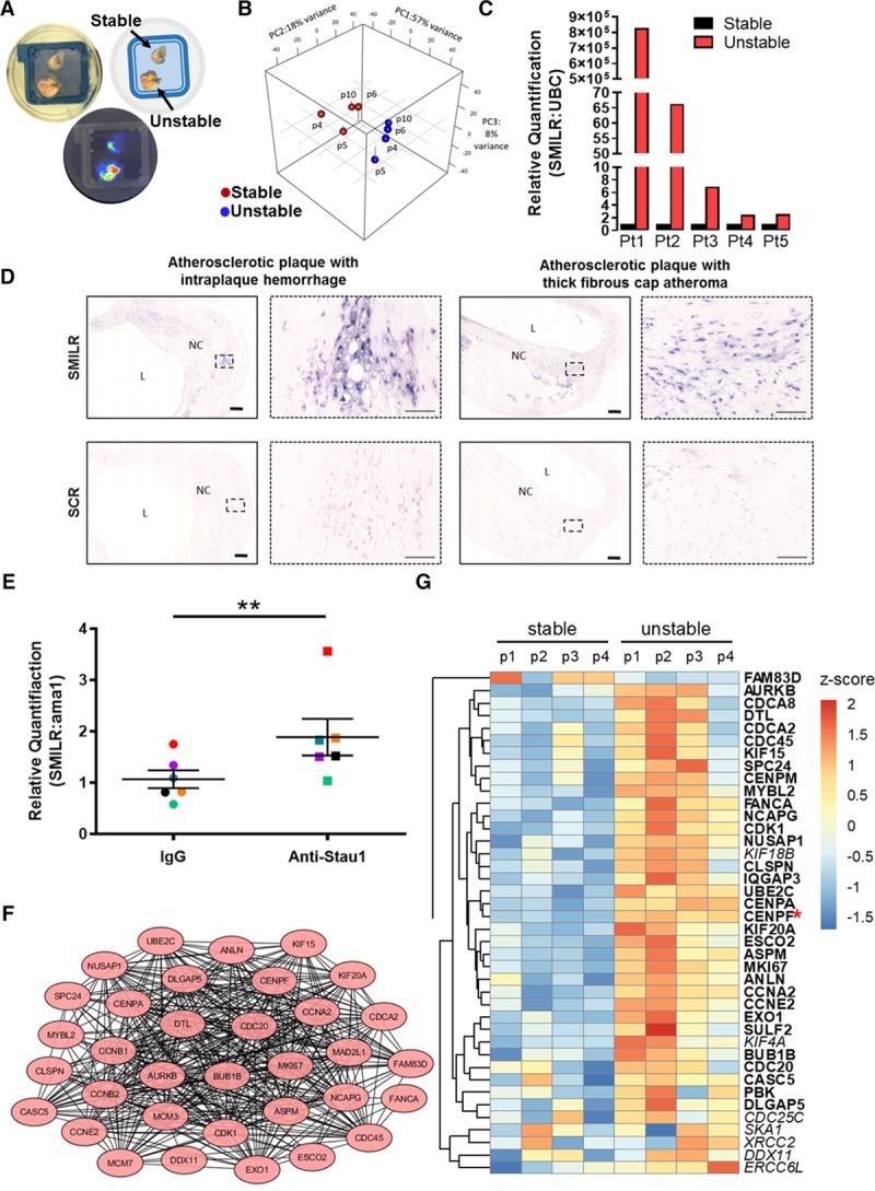
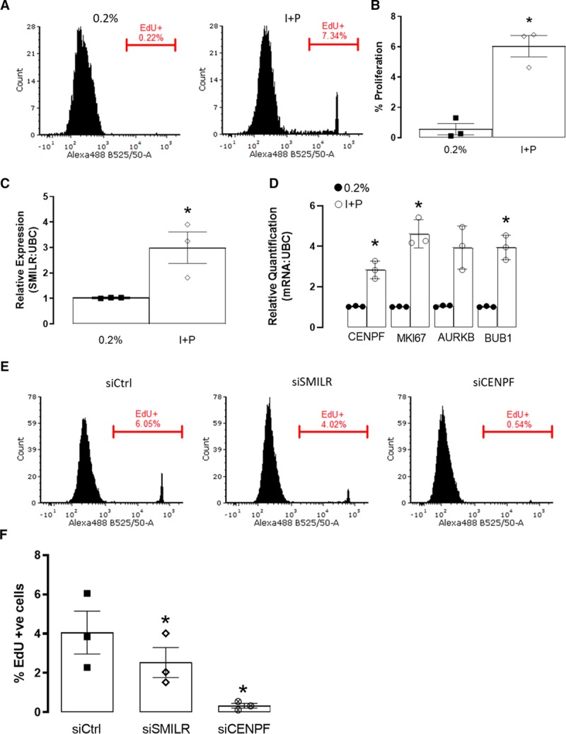
We used deep RNA-sequencing of human saphenous vein SMCs stimulated with IL (interleukin)-1α and PDGF (platelet-derived growth factor)-BB with SMILR knockdown (siRNA) or overexpression (lentivirus), to identify SMILR-regulated genes. This revealed a SMILR-dependent network essential for cell cycle progression. In particular, we found using the fluorescent ubiquitination-based cell cycle indicator viral system that SMILR regulates the late mitotic phase of the cell cycle and cytokinesis with SMILR knockdown resulting in ≈10% increase in binucleated cells. SMILR pulldowns further revealed its potential molecular mechanism, which involves an interaction with the mRNA of the late mitotic protein CENPF (centromere protein F) and the regulatory Staufen1 RNA-binding protein. SMILR and this downstream axis were also found to be activated in the human ex vivo vein graft pathological model and in primary human coronary artery SMCs and atherosclerotic plaques obtained at carotid endarterectomy. Finally, to assess the therapeutic potential of SMILR, we used a novel siRNA approach in the ex vivo vein graft model (within the 30 minutes clinical time frame that would occur between harvest and implant) to assess the reduction of proliferation by EdU incorporation. SMILR knockdown led to a marked decrease in proliferation from ≈29% in controls to ≈5% with SMILR depletion.
Collectively, we demonstrate that SMILR is a critical mediator of vascular SMC proliferation via direct regulation of mitotic progression. Our data further reveal a potential SMILR-targeting intervention to limit atherogenesis and adverse vascular remodeling.
血管壁损伤后,血管平滑肌细胞(VSMC)异常增殖导致病理性重塑。然而,其调控机制尚不完全清楚。
我们最近发现,人类长非编码 RNA SMILR 通过一种未知的机制促进血管平滑肌细胞增殖。在此,我们评估了 SMILR 抑制的治疗潜力,并详细阐述了其作用机制。
我们采用 IL-1α和 PDGF-BB 刺激的人隐静脉平滑肌细胞的深度 RNA 测序,用 SMILR 敲低(siRNA)或过表达(慢病毒)处理,鉴定 SMILR 调控的基因。这揭示了一个对细胞周期进程至关重要的 SMILR 依赖网络。特别是,我们发现,使用荧光泛素化细胞周期指示剂病毒系统,SMILR 调控细胞周期的晚期有丝分裂阶段和胞质分裂,SMILR 敲低导致双核细胞增加约 10%。SMILR 下拉实验进一步揭示了其潜在的分子机制,涉及与晚期有丝分裂蛋白 CENPF(着丝粒蛋白 F)mRNA和调节蛋白 Staufen1 RNA 结合蛋白的相互作用。SMILR 及其下游轴在人类动静脉移植物病理模型和颈动脉内膜切除术获得的原代人冠状动脉平滑肌细胞和动脉粥样硬化斑块中也被发现被激活。最后,为了评估 SMILR 的治疗潜力,我们在动静脉移植物模型中使用了一种新的 siRNA 方法(在收获和植入之间发生的 30 分钟临床时间范围内),通过 EdU 掺入评估增殖减少情况。SMILR 敲低导致增殖从对照组的约 29%显著降低至 SMILR 耗竭组的约 5%。
总之,我们证明 SMILR 是血管平滑肌细胞增殖的关键介质,通过直接调节有丝分裂进程。我们的数据进一步揭示了一种潜在的 SMILR 靶向干预方法,以限制动脉粥样硬化形成和血管不良重塑。