Department of Public Health and Caring Sciences/Geriatrics, Uppsala University, Rudbeck Laboratory, 75185, Uppsala, Sweden.
Eur J Nucl Med Mol Imaging. 2019 Dec;46(13):2848-2858. doi: 10.1007/s00259-019-04426-0. Epub 2019 Jul 24.
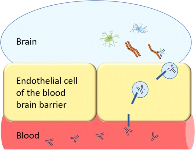
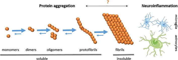
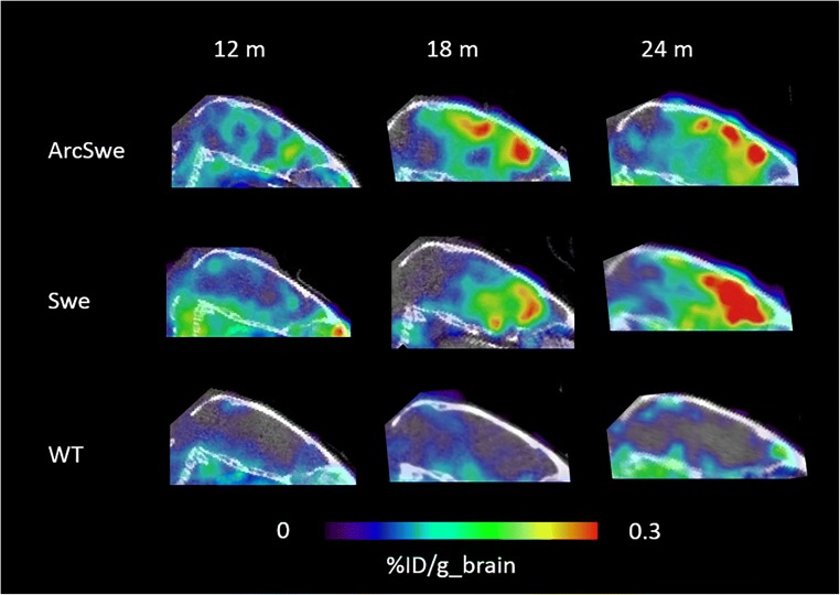
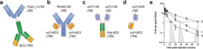
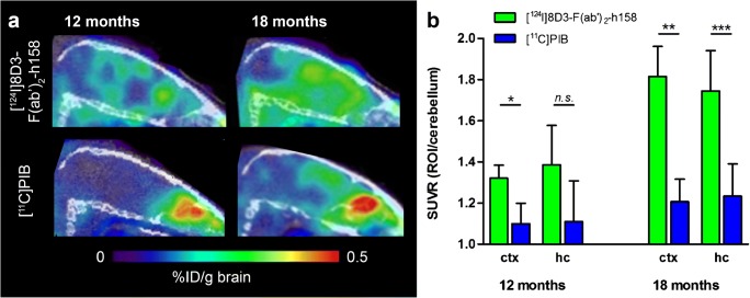
Almost 50 million people worldwide are affected by Alzheimer's disease (AD), the most common neurodegenerative disorder. Development of disease-modifying therapies would benefit from reliable, non-invasive positron emission tomography (PET) biomarkers for early diagnosis, monitoring of disease progression, and assessment of therapeutic effects. Traditionally, PET ligands have been based on small molecules that, with the right properties, can penetrate the blood-brain barrier (BBB) and visualize targets in the brain. Recently a new class of PET ligands based on antibodies have emerged, mainly in applications related to cancer. While antibodies have advantages such as high specificity and affinity, their passage across the BBB is limited. Thus, to be used as brain PET ligands, antibodies need to be modified for active transport into the brain. Here, we review the development of radioligands based on antibodies for visualization of intrabrain targets. We focus on antibodies modified into a bispecific format, with the capacity to undergo transferrin receptor 1 (TfR1)-mediated transcytosis to enter the brain and access pathological proteins, e.g. amyloid-beta. A number of such antibody ligands have been developed, displaying differences in brain uptake, pharmacokinetics, and ability to bind and visualize the target in the brain of transgenic mice. Potential pathological changes related to neurodegeneration, e.g. misfolded proteins and neuroinflammation, are suggested as future targets for this novel type of radioligand. Challenges are also discussed, such as the temporal match of radionuclide half-life with the ligand's pharmacokinetic profile and translation to human use. In conclusion, brain PET imaging using bispecific antibodies, modified for receptor-mediated transcytosis across the BBB, is a promising method for specifically visualizing molecules in the brain that are difficult to target with traditional small molecule ligands.
全球有近 5000 万人受到阿尔茨海默病(AD)的影响,这是最常见的神经退行性疾病。开发疾病修饰疗法将受益于可靠的、非侵入性的正电子发射断层扫描(PET)生物标志物,用于早期诊断、疾病进展监测和治疗效果评估。传统上,PET 配体基于小分子,这些小分子具有适当的特性,可以穿透血脑屏障(BBB)并可视化大脑中的靶标。最近,出现了一类基于抗体的新型 PET 配体,主要应用于癌症相关领域。虽然抗体具有高特异性和亲和力等优点,但它们穿过 BBB 的能力有限。因此,要将其用作脑 PET 配体,需要对抗体进行修饰以实现主动转运进入大脑。在这里,我们综述了基于抗体的放射性配体用于可视化脑内靶标的开发。我们重点介绍了修饰为双特异性形式的抗体,具有通过转铁蛋白受体 1(TfR1)介导的转胞吞作用进入大脑并与病理性蛋白(例如淀粉样β)结合的能力。已经开发了许多此类抗体配体,它们在脑摄取、药代动力学和在转基因小鼠大脑中结合和可视化靶标的能力方面存在差异。与神经退行性变相关的潜在病理变化,例如错误折叠的蛋白质和神经炎症,被认为是这种新型放射性配体的未来靶标。还讨论了一些挑战,例如放射性核素半衰期与配体药代动力学特征的时间匹配以及向人类应用的转化。总之,用于受体介导的跨 BBB 转胞吞作用的双特异性抗体脑 PET 成像,是一种有前途的方法,可特异性地可视化用传统小分子配体难以靶向的大脑内分子。