Pulmonary Service, Clínica Universidad de Navarra, Pamplona, Spain.
Department of Radiology Mount Sinai School of Medicine, NY, United States of America.
PLoS One. 2019 Jul 25;14(7):e0219187. doi: 10.1371/journal.pone.0219187. eCollection 2019.
To assess the relationship between lung cancer and emphysema subtypes.
Airflow obstruction and emphysema predispose to lung cancer. Little is known, however, about the lung cancer risk associated with different emphysema phenotypes. We assessed the risk of lung cancer based on the presence, type and severity of emphysema, using visual assessment.
Seventy-two consecutive lung cancer cases were selected from a prospective cohort of 3,477 participants enrolled in the Clínica Universidad de Navarra's lung cancer screening program. Each case was matched to three control subjects using age, sex, smoking history and body mass index as key variables. Visual assessment of emphysema and spirometry were performed. Logistic regression and interaction model analysis were used in order to investigate associations between lung cancer and emphysema subtypes.
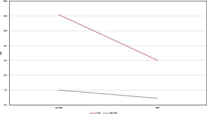
Airflow obstruction and visual emphysema were significantly associated with lung cancer (OR = 2.8, 95%CI: 1.6 to 5.2; OR = 5.9, 95%CI: 2.9 to 12.2; respectively). Emphysema severity and centrilobular subtype were associated with greater risk when adjusted for confounders (OR = 12.6, 95%CI: 1.6 to 99.9; OR = 34.3, 95%CI: 25.5 to 99.3, respectively). The risk of lung cancer decreases with the added presence of paraseptal emphysema (OR = 4.0, 95%CI: 3.6 to 34.9), losing this increased risk of lung cancer when it occurs alone (OR = 0.7, 95%CI: 0.5 to 2.6).
Visual scoring of emphysema predicts lung cancer risk. The centrilobular phenotype is associated with the greatest risk.
评估肺癌与肺气肿亚型之间的关系。
气流阻塞和肺气肿易导致肺癌。然而,对于不同肺气肿表型与肺癌风险的关系知之甚少。我们通过视觉评估,根据肺气肿的存在、类型和严重程度来评估肺癌的风险。
从在纳瓦拉大学临床医院肺癌筛查计划中纳入的 3477 名参与者的前瞻性队列中选择了 72 例连续肺癌病例。每个病例都通过年龄、性别、吸烟史和体重指数等关键变量与 3 个对照进行匹配。进行了肺气肿的视觉评估和肺量测定。使用逻辑回归和交互模型分析来研究肺癌与肺气肿亚型之间的关联。
气流阻塞和视觉肺气肿与肺癌显著相关(OR = 2.8,95%CI:1.6 至 5.2;OR = 5.9,95%CI:2.9 至 12.2;分别)。在校正混杂因素后,肺气肿严重程度和中央型亚型与更高的风险相关(OR = 12.6,95%CI:1.6 至 99.9;OR = 34.3,95%CI:25.5 至 99.3,分别)。当存在旁间隔肺气肿时,肺癌风险降低(OR = 4.0,95%CI:3.6 至 34.9),当单独存在时,肺癌风险降低(OR = 0.7,95%CI:0.5 至 2.6)。
肺气肿的视觉评分可预测肺癌风险。中央型表型与最大风险相关。