• 文献检索
  • 文档翻译
  • 深度研究
  • 学术资讯
  • Suppr Zotero 插件Zotero 插件
  • 邀请有礼
  • 套餐&价格
  • 历史记录
应用&插件
Suppr Zotero 插件Zotero 插件浏览器插件Mac 客户端Windows 客户端微信小程序
定价
高级版会员购买积分包购买API积分包
服务
文献检索文档翻译深度研究API 文档MCP 服务
关于我们
关于 Suppr公司介绍联系我们用户协议隐私条款
关注我们

Suppr 超能文献

核心技术专利:CN118964589B侵权必究
粤ICP备2023148730 号-1Suppr @ 2026

文献检索

告别复杂PubMed语法,用中文像聊天一样搜索,搜遍4000万医学文献。AI智能推荐,让科研检索更轻松。

立即免费搜索

文件翻译

保留排版,准确专业,支持PDF/Word/PPT等文件格式,支持 12+语言互译。

免费翻译文档

深度研究

AI帮你快速写综述,25分钟生成高质量综述,智能提取关键信息,辅助科研写作。

立即免费体验

基于注意力的多实例深度学习在胸部低剂量 CT 肺气肿评估中的应用。

Evaluation of emphysema on thoracic low-dose CTs through attention-based multiple instance deep learning.

机构信息

Committee on Medical Physics, Department of Radiology, The University of Chicago, 5841 S Maryland Avenue, MC2026, Chicago, 60637, USA.

Department of Radiology, Icahn School of Medicine at Mount Sinai, New York, 10029, USA.

出版信息

Sci Rep. 2023 Jan 21;13(1):1187. doi: 10.1038/s41598-023-27549-9.

DOI:10.1038/s41598-023-27549-9
PMID:36681685
原文链接:https://pmc.ncbi.nlm.nih.gov/articles/PMC9867724/
Abstract

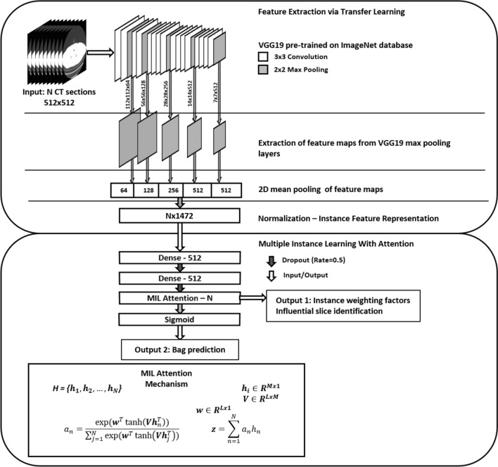
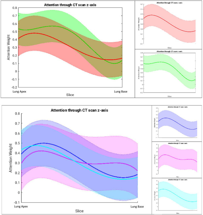
In addition to lung cancer, other thoracic abnormalities, such as emphysema, can be visualized within low-dose CT scans that were initially obtained in cancer screening programs, and thus, opportunistic evaluation of these diseases may be highly valuable. However, manual assessment for each scan is tedious and often subjective, thus we have developed an automatic, rapid computer-aided diagnosis system for emphysema using attention-based multiple instance deep learning and 865 LDCTs. In the task of determining if a CT scan presented with emphysema or not, our novel Transfer AMIL approach yielded an area under the ROC curve of 0.94 ± 0.04, which was a statistically significant improvement compared to other methods evaluated in our study following the Delong Test with correction for multiple comparisons. Further, from our novel attention weight curves, we found that the upper lung demonstrated a stronger influence in all scan classes, indicating that the model prioritized upper lobe information. Overall, our novel Transfer AMIL method yielded high performance and provided interpretable information by identifying slices that were most influential to the classification decision, thus demonstrating strong potential for clinical implementation.

摘要

除了肺癌,在最初用于癌症筛查计划的低剂量 CT 扫描中,还可以观察到其他胸部异常,如肺气肿。因此,对这些疾病进行机会性评估可能具有很高的价值。然而,对每个扫描进行手动评估既繁琐又主观,因此我们已经开发了一种使用基于注意力的多实例深度学习和 865 个 LDCT 的肺气肿自动快速计算机辅助诊断系统。在确定 CT 扫描是否存在肺气肿的任务中,我们的新型 Transfer AMIL 方法在 ROC 曲线下的面积为 0.94 ± 0.04,与我们在研究中使用 Delong 测试校正多重比较评估的其他方法相比,这是一个具有统计学意义的改进。此外,从我们的新型注意力权重曲线中,我们发现上肺在所有扫描类别中都表现出更强的影响,这表明该模型优先考虑上叶信息。总体而言,我们的新型 Transfer AMIL 方法具有出色的性能,并通过识别对分类决策最有影响的切片来提供可解释的信息,因此具有很强的临床应用潜力。

https://cdn.ncbi.nlm.nih.gov/pmc/blobs/3559/9867724/58f54f28974d/41598_2023_27549_Fig3_HTML.jpg
https://cdn.ncbi.nlm.nih.gov/pmc/blobs/3559/9867724/0678a031403e/41598_2023_27549_Fig1_HTML.jpg
https://cdn.ncbi.nlm.nih.gov/pmc/blobs/3559/9867724/17d70c5b7ab5/41598_2023_27549_Fig2_HTML.jpg
https://cdn.ncbi.nlm.nih.gov/pmc/blobs/3559/9867724/58f54f28974d/41598_2023_27549_Fig3_HTML.jpg
https://cdn.ncbi.nlm.nih.gov/pmc/blobs/3559/9867724/0678a031403e/41598_2023_27549_Fig1_HTML.jpg
https://cdn.ncbi.nlm.nih.gov/pmc/blobs/3559/9867724/17d70c5b7ab5/41598_2023_27549_Fig2_HTML.jpg
https://cdn.ncbi.nlm.nih.gov/pmc/blobs/3559/9867724/58f54f28974d/41598_2023_27549_Fig3_HTML.jpg

相似文献

1
Evaluation of emphysema on thoracic low-dose CTs through attention-based multiple instance deep learning.基于注意力的多实例深度学习在胸部低剂量 CT 肺气肿评估中的应用。
Sci Rep. 2023 Jan 21;13(1):1187. doi: 10.1038/s41598-023-27549-9.
2
Deep Learning-Based Kernel Adaptation Enhances Quantification of Emphysema on Low-Dose Chest CT for Predicting Long-Term Mortality.基于深度学习的核适应增强了低剂量胸部CT上肺气肿的量化以预测长期死亡率。
Invest Radiol. 2024 Mar 1;59(3):278-286. doi: 10.1097/RLI.0000000000001003.
3
Emphysema quantification using low-dose computed tomography with deep learning-based kernel conversion comparison.使用基于深度学习的核转换比较的低剂量 CT 进行肺气肿定量。
Eur Radiol. 2020 Dec;30(12):6779-6787. doi: 10.1007/s00330-020-07020-3. Epub 2020 Jun 29.
4
Deep learning-enabled accurate normalization of reconstruction kernel effects on emphysema quantification in low-dose CT.深度学习可实现低剂量 CT 肺气肿定量中重建核效应的准确归一化。
Phys Med Biol. 2019 Jul 4;64(13):135010. doi: 10.1088/1361-6560/ab28a1.
5
AI-Driven Model for Automatic Emphysema Detection in Low-Dose Computed Tomography Using Disease-Specific Augmentation.基于疾病特异性增强的低剂量 CT 自动肺气肿检测的人工智能模型
J Digit Imaging. 2022 Jun;35(3):538-550. doi: 10.1007/s10278-022-00599-7. Epub 2022 Feb 18.
6
Emphysema Quantification Using Ultra-Low-Dose Chest CT: Efficacy of Deep Learning-Based Image Reconstruction.使用超低剂量胸部 CT 进行肺气肿定量:基于深度学习的图像重建的效果。
Medicina (Kaunas). 2022 Jul 15;58(7):939. doi: 10.3390/medicina58070939.
7
Automatic emphysema detection using weakly labeled HRCT lung images.利用弱标注 HRCT 肺图像进行自动肺气肿检测。
PLoS One. 2018 Oct 15;13(10):e0205397. doi: 10.1371/journal.pone.0205397. eCollection 2018.
8
A convolutional neural network for ultra-low-dose CT denoising and emphysema screening.用于超低剂量 CT 去噪和肺气肿筛查的卷积神经网络。
Med Phys. 2019 Sep;46(9):3941-3950. doi: 10.1002/mp.13666. Epub 2019 Jul 17.
9
Pulmonary emphysema quantification at low dose chest CT using Deep Learning image reconstruction.低剂量胸部 CT 应用深度学习图像重建对肺气肿进行定量分析。
Eur J Radiol. 2022 Jul;152:110338. doi: 10.1016/j.ejrad.2022.110338. Epub 2022 May 5.
10
A homological approach to a mathematical definition of pulmonary fibrosis and emphysema on computed tomography.一种基于同调理论的 CT 图像中肺纤维化和肺气肿的数学定义方法。
J Appl Physiol (1985). 2021 Aug 1;131(2):601-612. doi: 10.1152/japplphysiol.00150.2021. Epub 2021 Jun 17.

引用本文的文献

1
Applying Deep Transfer Learning to Assess the Impact of Imaging Modalities on Colon Cancer Detection.应用深度迁移学习评估成像模态对结肠癌检测的影响。
Diagnostics (Basel). 2023 May 12;13(10):1721. doi: 10.3390/diagnostics13101721.
2
Clinical Scores, Biomarkers and IT Tools in Lung Cancer Screening-Can an Integrated Approach Overcome Current Challenges?肺癌筛查中的临床评分、生物标志物和信息技术工具——综合方法能否克服当前挑战?
Cancers (Basel). 2023 Feb 14;15(4):1218. doi: 10.3390/cancers15041218.

本文引用的文献

1
Emphysema Progression at CT by Deep Learning Predicts Functional Impairment and Mortality: Results from the COPDGene Study.深度学习预测 CT 肺气肿进展与功能障碍和死亡率的关系:COPDGene 研究结果。
Radiology. 2022 Sep;304(3):672-679. doi: 10.1148/radiol.213054. Epub 2022 May 17.
2
Value-added Opportunistic CT Screening: State of the Art.增值性机会性 CT 筛查:现状。
Radiology. 2022 May;303(2):241-254. doi: 10.1148/radiol.211561. Epub 2022 Mar 15.
3
The importance of low-dose CT screening to identify emphysema in asymptomatic participants with and without a prior diagnosis of COPD.
低剂量 CT 筛查在有和无 COPD 既往诊断的无症状参与者中识别肺气肿的重要性。
Clin Imaging. 2021 Oct;78:136-141. doi: 10.1016/j.clinimag.2021.03.012. Epub 2021 Mar 20.
4
Added benefits of early detection of other diseases on low-dose CT screening.低剂量CT筛查时早期发现其他疾病的额外益处。
Transl Lung Cancer Res. 2021 Feb;10(2):1141-1153. doi: 10.21037/tlcr-20-746.
5
Screening for Lung Cancer: US Preventive Services Task Force Recommendation Statement.肺癌筛查:美国预防服务工作组推荐声明。
JAMA. 2021 Mar 9;325(10):962-970. doi: 10.1001/jama.2021.1117.
6
HRCT characteristics of severe emphysema patients: Interobserver variability among expert readers and comparison with quantitative software.严重肺气肿患者的 HRCT 特征:专家读者之间的观察者间变异性及与定量软件的比较。
Eur J Radiol. 2021 Mar;136:109561. doi: 10.1016/j.ejrad.2021.109561. Epub 2021 Jan 23.
7
Quantitative Emphysema on Low-Dose CT Imaging of the Chest and Risk of Lung Cancer and Airflow Obstruction: An Analysis of the National Lung Screening Trial.胸部低剂量 CT 成像定量肺气肿与肺癌及气流阻塞风险:国家肺癌筛查试验分析。
Chest. 2021 May;159(5):1812-1820. doi: 10.1016/j.chest.2020.12.004. Epub 2020 Dec 14.
8
An End-to-End Deep Learning Pipeline for Emphysema Quantification Using Multi-label Learning.一种使用多标签学习进行肺气肿定量分析的端到端深度学习管道。
Annu Int Conf IEEE Eng Med Biol Soc. 2019 Jul;2019:929-932. doi: 10.1109/EMBC.2019.8857392.
9
Deep Learning Enables Automatic Classification of Emphysema Pattern at CT.深度学习可实现 CT 肺气肿模式的自动分类。
Radiology. 2020 Feb;294(2):434-444. doi: 10.1148/radiol.2019191022. Epub 2019 Dec 3.
10
Classification of Volumetric Images Using Multi-Instance Learning and Extreme Value Theorem.基于多实例学习和极值定理的体绘制图像分类。
IEEE Trans Med Imaging. 2020 Apr;39(4):854-865. doi: 10.1109/TMI.2019.2936244. Epub 2019 Aug 19.