• 文献检索
  • 文档翻译
  • 深度研究
  • 学术资讯
  • Suppr Zotero 插件Zotero 插件
  • 邀请有礼
  • 套餐&价格
  • 历史记录
应用&插件
Suppr Zotero 插件Zotero 插件浏览器插件Mac 客户端Windows 客户端微信小程序
定价
高级版会员购买积分包购买API积分包
服务
文献检索文档翻译深度研究API 文档MCP 服务
关于我们
关于 Suppr公司介绍联系我们用户协议隐私条款
关注我们

Suppr 超能文献

核心技术专利:CN118964589B侵权必究
粤ICP备2023148730 号-1Suppr @ 2026

文献检索

告别复杂PubMed语法,用中文像聊天一样搜索,搜遍4000万医学文献。AI智能推荐,让科研检索更轻松。

立即免费搜索

文件翻译

保留排版,准确专业,支持PDF/Word/PPT等文件格式,支持 12+语言互译。

免费翻译文档

深度研究

AI帮你快速写综述,25分钟生成高质量综述,智能提取关键信息,辅助科研写作。

立即免费体验

慢性中心性浆液性脉络膜视网膜病变消退后的视力和视网膜形态损害

Impairment of visual acuity and retinal morphology following resolved chronic central serous chorioretinopathy.

作者信息

Gawęcki Maciej, Jaszczuk-Maciejewska Agnieszka, Jurska-Jaśko Anna, Kneba Małgorzata, Grzybowski Andrzej

机构信息

Dobry Wzrok Ophthalmological Clinic, Gdańsk, Poland.

Department of Ophthalmology, Univeristy of Warmia and Mazury, Olsztyn, Poland.

出版信息

BMC Ophthalmol. 2019 Jul 25;19(1):160. doi: 10.1186/s12886-019-1171-5.

DOI:10.1186/s12886-019-1171-5
PMID:31345183
原文链接:https://pmc.ncbi.nlm.nih.gov/articles/PMC6659242/
Abstract

PURPOSE

Central serous chorioretinopathy (CSCR) is a complex ocular entity that, in its chronic form, can lead to serious visual impairment and morphological damage to the retina. The aim of the current retrospective study was to evaluate the damage present after long-standing but resolved central serous chorioretinopathy and refer it to healthy individuals. Correlations between measurable factors-for example, duration of the disease, baseline retinal morphological parameters, or patient age and/or their degree of impairment-were also assessed.

MATERIALS AND METHODS

The study group consisted of thirty-two eyes (13 female and 19 male, mean age 49.6 years SD +/- 10.5) with chronic central serous chorioretinopathy (mean duration 18.9 months SD +/- 15.4) in which complete resolution of subretinal fluid was achieved after subthreshold micropulse laser treatment. Inclusion criterion was a lack of subretinal fluid within the whole area of the central retina scanned by the spectral domain optical coherence tomography. The group was extracted out of 51 cases of chronic CSCR that were treated with that method. They were analyzed according to final best-corrected visual acuity and retinal morphological parameters as measured by spectral optical coherence tomography with angiography option (OCTA). Results were compared with the outcomes of a control group, which consisted of 40 eyes of healthy individuals with full distance visual acuity (0.0 logMAR, 1.0 Snellen) never treated with subthreshold micropulse laser. Statistical analysis included regarding correlation between final visual acuity and final central retinal thickness and retinal and functional parameters prior to treatment.

RESULTS

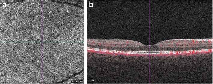
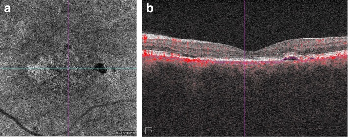
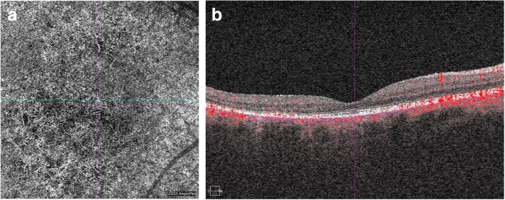
Final best-corrected visual acuity after chronic central serous chorioretinopathy was 0.23 logMAR (0.6 Snellen) and central retinal thickness was 39.32 μm smaller than in controls. No correlation was found between final visual acuity and retinal thickness and duration of the disease, patient age, and baseline morphological retinal parameters. OCTA scans revealed impaired choriocapillaries flow signal even following resolution of the disease.

CONCLUSION

Chronic central serous chorioretinopathy is a potentially damaging clinical entity that results in serious visual impairment, retinal thinning, and choroidal flow defects. Further research is needed to determine precisely the timepoint of this damage.

摘要

目的

中心性浆液性脉络膜视网膜病变(CSCR)是一种复杂的眼部疾病,其慢性形式可导致严重的视力损害和视网膜形态损伤。本项回顾性研究的目的是评估长期但已治愈的中心性浆液性脉络膜视网膜病变后存在的损伤,并将其与健康个体进行对比。还评估了可测量因素之间的相关性,例如疾病持续时间、基线视网膜形态参数、患者年龄和/或其损伤程度。

材料与方法

研究组由32只眼(13名女性和19名男性,平均年龄49.6岁,标准差±10.5)组成,患有慢性中心性浆液性脉络膜视网膜病变(平均病程18.9个月,标准差±15.4),经阈下微脉冲激光治疗后视网膜下液完全消退。纳入标准是在光谱域光学相干断层扫描所扫描的整个中央视网膜区域内不存在视网膜下液。该组是从51例采用该方法治疗的慢性CSCR病例中提取出来的。根据最终最佳矫正视力和通过具有血管造影选项的光谱光学相干断层扫描(OCTA)测量的视网膜形态参数对他们进行分析。将结果与对照组的结果进行比较,对照组由40只健康个体的眼睛组成,这些个体具有全距视力(0.0 logMAR,1.0 Snellen),从未接受过阈下微脉冲激光治疗。统计分析包括治疗前最终视力与最终中央视网膜厚度以及视网膜和功能参数之间的相关性。

结果

慢性中心性浆液性脉络膜视网膜病变后的最终最佳矫正视力为0.23 logMAR(0.6 Snellen),中央视网膜厚度比对照组小39.32μm。在最终视力与视网膜厚度、疾病持续时间、患者年龄和基线视网膜形态参数之间未发现相关性。OCTA扫描显示即使在疾病消退后脉络膜毛细血管血流信号仍受损。

结论

慢性中心性浆液性脉络膜视网膜病变是一种潜在的破坏性临床疾病,可导致严重的视力损害、视网膜变薄和脉络膜血流缺陷。需要进一步研究以精确确定这种损伤的时间点。

https://cdn.ncbi.nlm.nih.gov/pmc/blobs/c8b0/6659242/78e0466569ed/12886_2019_1171_Fig3_HTML.jpg
https://cdn.ncbi.nlm.nih.gov/pmc/blobs/c8b0/6659242/2f96ca0bbccb/12886_2019_1171_Fig1_HTML.jpg
https://cdn.ncbi.nlm.nih.gov/pmc/blobs/c8b0/6659242/73dcf0ef18b1/12886_2019_1171_Fig2_HTML.jpg
https://cdn.ncbi.nlm.nih.gov/pmc/blobs/c8b0/6659242/78e0466569ed/12886_2019_1171_Fig3_HTML.jpg
https://cdn.ncbi.nlm.nih.gov/pmc/blobs/c8b0/6659242/2f96ca0bbccb/12886_2019_1171_Fig1_HTML.jpg
https://cdn.ncbi.nlm.nih.gov/pmc/blobs/c8b0/6659242/73dcf0ef18b1/12886_2019_1171_Fig2_HTML.jpg
https://cdn.ncbi.nlm.nih.gov/pmc/blobs/c8b0/6659242/78e0466569ed/12886_2019_1171_Fig3_HTML.jpg

相似文献

1
Impairment of visual acuity and retinal morphology following resolved chronic central serous chorioretinopathy.慢性中心性浆液性脉络膜视网膜病变消退后的视力和视网膜形态损害
BMC Ophthalmol. 2019 Jul 25;19(1):160. doi: 10.1186/s12886-019-1171-5.
2
Functional and morphological outcome in patients with chronic central serous chorioretinopathy treated by subthreshold micropulse laser.经阈下微脉冲激光治疗的慢性中心性浆液性脉络膜视网膜病变患者的功能和形态学转归
Graefes Arch Clin Exp Ophthalmol. 2017 Dec;255(12):2299-2306. doi: 10.1007/s00417-017-3783-x. Epub 2017 Aug 22.
3
Clinical evaluation of neovascular and non-neovascular chronic central serous chorioretinopathy (CSC) diagnosed by swept source optical coherence tomography angiography (SS OCTA).经扫频源光学相干断层扫描血管造影(SS OCTA)诊断的新生血管性和非新生血管性慢性中心性浆液性脉络膜视网膜病变(CSC)的临床评估
Graefes Arch Clin Exp Ophthalmol. 2019 Aug;257(8):1581-1590. doi: 10.1007/s00417-019-04297-z. Epub 2019 Apr 29.
4
BASELINE SPECTRAL DOMAIN OPTICAL COHERENCE TOMOGRAPHIC HYPERREFLECTIVE FOCI AS A PREDICTOR OF VISUAL OUTCOME AND RECURRENCE FOR CENTRAL SEROUS CHORIORETINOPATHY.基线光谱域光学相干断层扫描高反射灶作为中心性浆液性脉络膜视网膜病变视觉预后和复发的预测指标
Retina. 2016 Jul;36(7):1372-80. doi: 10.1097/IAE.0000000000000929.
5
Photodynamic therapy for chronic central serous chorioretinopathy: a 4-year follow-up study.光动力疗法治疗慢性中心性浆液性脉络膜视网膜病变:4 年随访研究。
Retina. 2013 Feb;33(2):309-15. doi: 10.1097/IAE.0b013e3182670fbe.
6
Ganglion Cell Loss in the Course of Central Serous Chorioretinopathy.中心性浆液性脉络膜视网膜病变病程中的神经节细胞丢失
Ophthalmol Ther. 2023 Feb;12(1):517-533. doi: 10.1007/s40123-022-00625-5. Epub 2022 Dec 12.
7
Retinal sensitivity after selective retina therapy (SRT) on patients with central serous chorioretinopathy.中心性浆液性脉络膜视网膜病变患者接受选择性视网膜治疗(SRT)后的视网膜敏感性
Graefes Arch Clin Exp Ophthalmol. 2017 Feb;255(2):243-254. doi: 10.1007/s00417-016-3441-8. Epub 2016 Aug 6.
8
The efficacy of subthreshold micropulse yellow laser (577 nm) in chronic central serous chorioretinopathy.阈下微脉冲黄色激光(577nm)治疗慢性中心性浆液性脉络膜视网膜病变的疗效。
Lasers Med Sci. 2021 Jul;36(5):981-988. doi: 10.1007/s10103-020-03129-5. Epub 2020 Aug 19.
9
Oral Rifampin treatment for longstanding chronic central serous chorioretinopathy.口服利福平治疗长期慢性中心性浆液性脉络膜视网膜病变。
Graefes Arch Clin Exp Ophthalmol. 2016 Jan;254(1):15-22. doi: 10.1007/s00417-015-2989-z. Epub 2015 Mar 21.
10
Half-Fluence Photodynamic Therapy for Chronic Central Serous Chorioretinopathy: Predisposing Factors for Visual Acuity Outcomes.半剂量光动力疗法治疗慢性中心性浆液性脉络膜视网膜病变:视力预后的易感因素
Semin Ophthalmol. 2018;33(5):690-699. doi: 10.1080/08820538.2017.1416414. Epub 2017 Dec 18.

引用本文的文献

1
Comparison of microvasculature between neovascular, non-neovascular chronic central serous chorioretinopathy cases and asymptomatic fellow eyes using swept-source optical coherence tomography angiography.使用扫频光学相干断层扫描血管造影术比较新生血管性、非新生血管性慢性中心性浆液性脉络膜视网膜病变病例与无症状对侧眼之间的微血管系统。
Int Ophthalmol. 2025 Jul 18;45(1):297. doi: 10.1007/s10792-025-03673-5.
2
Predictive Factors for Morphological and Functional Improvements in Long-Lasting Central Serous Chorioretinopathy Treated with Photodynamic Therapy.光动力疗法治疗慢性中心性浆液性脉络膜视网膜病变形态学和功能改善的预测因素
Biomedicines. 2025 Apr 11;13(4):944. doi: 10.3390/biomedicines13040944.
3

本文引用的文献

1
Three-Year Results of Fluorescein Angiography-Guided Standard Photodynamic Therapy with Multiple Spots for Central Serous Chorioretinopathy.荧光素血管造影引导下多点标准光动力疗法治疗中心性浆液性脉络膜视网膜病变的三年结果
Ophthalmol Retina. 2018 Jul;2(7):703-711. doi: 10.1016/j.oret.2017.11.008. Epub 2017 Dec 29.
2
Mineralocorticoid Receptor Antagonists in Central Serous Chorioretinopathy: A Meta-Analysis of Randomized Controlled Trials.盐皮质激素受体拮抗剂治疗中心性浆液性脉络膜视网膜病变:一项随机对照试验的荟萃分析
Ophthalmol Retina. 2019 Feb;3(2):154-160. doi: 10.1016/j.oret.2018.09.003. Epub 2018 Sep 15.
3
Anatomical and Functional Changes of the Retina and the Choroid after Resolved Chronic CSCR.
Quantitative Changes in Vascular and Neural Fibers Induced by Subretinal Fluid Excluding the Peripapillary Region in Patients with Chronic Central Serous Chorioretinopathy.
慢性中心性浆液性脉络膜视网膜病变患者视网膜下液(不包括视乳头周围区域)诱导的血管和神经纤维的定量变化
Diagnostics (Basel). 2025 Jan 14;15(2):174. doi: 10.3390/diagnostics15020174.
4
The role of inflammation in central serous chorioretinopathy: From mechanisms to therapeutic prospects.炎症在中心性浆液性脉络膜视网膜病变中的作用:从机制到治疗前景
Front Pharmacol. 2024 May 21;15:1200492. doi: 10.3389/fphar.2024.1200492. eCollection 2024.
5
Quantification of Metamorphopsia in Resolved Idiopathic Central Serous Chorioretinopathy: An Analysis Using M-CHARTS, Amsler Grid, and Optical Coherence Tomography.已愈特发性中心性浆液性脉络膜视网膜病变的视物变形定量分析:使用M-CHARTS、阿姆斯勒方格表和光学相干断层扫描的分析
Clin Ophthalmol. 2024 Mar 27;18:937-942. doi: 10.2147/OPTH.S456556. eCollection 2024.
6
Crossover to PDT after the unsuccessful micropulse laser treatment of central serous chorioretinopathy.中心性浆液性脉络膜视网膜病变微脉冲激光治疗失败后交叉接受光动力疗法。
Adv Ophthalmol Pract Res. 2024 Jan 23;4(1):32-38. doi: 10.1016/j.aopr.2024.01.004. eCollection 2024 Feb-Mar.
7
Factors related to changes in visual symptoms after successful photodynamic therapy in central serous chorioretinopathy.与中心性浆液性脉络膜视网膜病变光动力疗法成功后视觉症状变化相关的因素。
PLoS One. 2023 Apr 21;18(4):e0284899. doi: 10.1371/journal.pone.0284899. eCollection 2023.
8
Biometric Risk Factors for Central Serous Chorioretinopathy.中心性浆液性脉络膜视网膜病变的生物统计学危险因素。
Ophthalmol Ther. 2023 Apr;12(2):1327-1338. doi: 10.1007/s40123-023-00687-z. Epub 2023 Feb 25.
9
Significant Visual Impairment after Short-Lasting Central Serous Chorioretinopathy.短期中心性浆液性脉络膜视网膜病变后的严重视力损害。
Case Rep Ophthalmol. 2022 Sep 19;13(3):678-685. doi: 10.1159/000525924. eCollection 2022 Fall.
10
Ganglion Cell Loss in the Course of Central Serous Chorioretinopathy.中心性浆液性脉络膜视网膜病变病程中的神经节细胞丢失
Ophthalmol Ther. 2023 Feb;12(1):517-533. doi: 10.1007/s40123-022-00625-5. Epub 2022 Dec 12.
慢性中心性浆液性脉络膜视网膜病变消退后视网膜和脉络膜的解剖及功能变化
J Clin Med. 2019 Apr 7;8(4):474. doi: 10.3390/jcm8040474.
4
A two-year study of diffused retinal pigment epitheliopathy treated with half-dose photodynamic therapy guided by simultaneous angiography and optical coherence tomography.一项为期两年的研究,对接受半剂量光动力疗法治疗的弥漫性视网膜色素上皮病变进行了同时的血管造影和光学相干断层扫描引导。
Eye (Lond). 2019 May;33(5):737-745. doi: 10.1038/s41433-018-0284-z. Epub 2018 Dec 7.
5
Clinical spectrum of severe chronic central serous chorioretinopathy and outcome of photodynamic therapy.重度慢性中心性浆液性脉络膜视网膜病变的临床谱及光动力疗法的疗效
Clin Ophthalmol. 2018 Oct 24;12:2167-2176. doi: 10.2147/OPTH.S174573. eCollection 2018.
6
Half-time photodynamic therapy in treatment of chronic central serous chorioretinopathy.半程光动力疗法治疗慢性中心性浆液性脉络膜视网膜病变
Graefes Arch Clin Exp Ophthalmol. 2018 Nov;256(11):2027-2034. doi: 10.1007/s00417-018-4086-6. Epub 2018 Aug 10.
7
Choroidal Vascular Reactivity in Central Serous Chorioretinopathy.脉络膜血管反应性在中心性浆液性脉络膜视网膜病变中的作用。
Invest Ophthalmol Vis Sci. 2018 Aug 1;59(10):3897-3905. doi: 10.1167/iovs.18-23995.
8
Subthreshold diode micropulse laser versus observation in acute central serous chorioretinopathy.阈下二极管微脉冲激光与急性中心性浆液性脉络膜视网膜病变观察的对比
Clin Exp Optom. 2019 Jan;102(1):79-85. doi: 10.1111/cxo.12818. Epub 2018 Jul 30.
9
Optical Coherence Tomography Angiography Quantitative Assessment of Choriocapillaris Blood Flow in Central Serous Chorioretinopathy.光学相干断层扫描血管造影定量评估中心性浆液性脉络膜视网膜病变脉络膜毛细血管血流。
Am J Ophthalmol. 2018 Oct;194:26-34. doi: 10.1016/j.ajo.2018.07.004. Epub 2018 Jul 24.
10
PATTERNS OF CHORIOCAPILLARIS FLOW SIGNAL VOIDS IN CENTRAL SEROUS CHORIORETINOPATHY: An Optical Coherence Tomography Angiography Study.脉络膜毛细血管血流信号缺失在中心性浆液性脉络膜视网膜病变中的表现:一项光学相干断层扫描血管造影研究。
Retina. 2019 Nov;39(11):2178-2188. doi: 10.1097/IAE.0000000000002271.