• 文献检索
  • 文档翻译
  • 深度研究
  • 学术资讯
  • Suppr Zotero 插件Zotero 插件
  • 邀请有礼
  • 套餐&价格
  • 历史记录
应用&插件
Suppr Zotero 插件Zotero 插件浏览器插件Mac 客户端Windows 客户端微信小程序
定价
高级版会员购买积分包购买API积分包
服务
文献检索文档翻译深度研究API 文档MCP 服务
关于我们
关于 Suppr公司介绍联系我们用户协议隐私条款
关注我们

Suppr 超能文献

核心技术专利:CN118964589B侵权必究
粤ICP备2023148730 号-1Suppr @ 2026

文献检索

告别复杂PubMed语法,用中文像聊天一样搜索,搜遍4000万医学文献。AI智能推荐,让科研检索更轻松。

立即免费搜索

文件翻译

保留排版,准确专业,支持PDF/Word/PPT等文件格式,支持 12+语言互译。

免费翻译文档

深度研究

AI帮你快速写综述,25分钟生成高质量综述,智能提取关键信息,辅助科研写作。

立即免费体验

基于深度学习方法对脑积水患儿进行自动心室系统分割。

Automated Ventricular System Segmentation in Paediatric Patients Treated for Hydrocephalus Using Deep Learning Methods.

机构信息

Department of Radiology, Poznań University of Medical Sciences, Poznań, Poland.

Fast-Radiology, Poland.

出版信息

Biomed Res Int. 2019 Jul 7;2019:3059170. doi: 10.1155/2019/3059170. eCollection 2019.

DOI:10.1155/2019/3059170
PMID:31360710
原文链接:https://pmc.ncbi.nlm.nih.gov/articles/PMC6642766/
Abstract

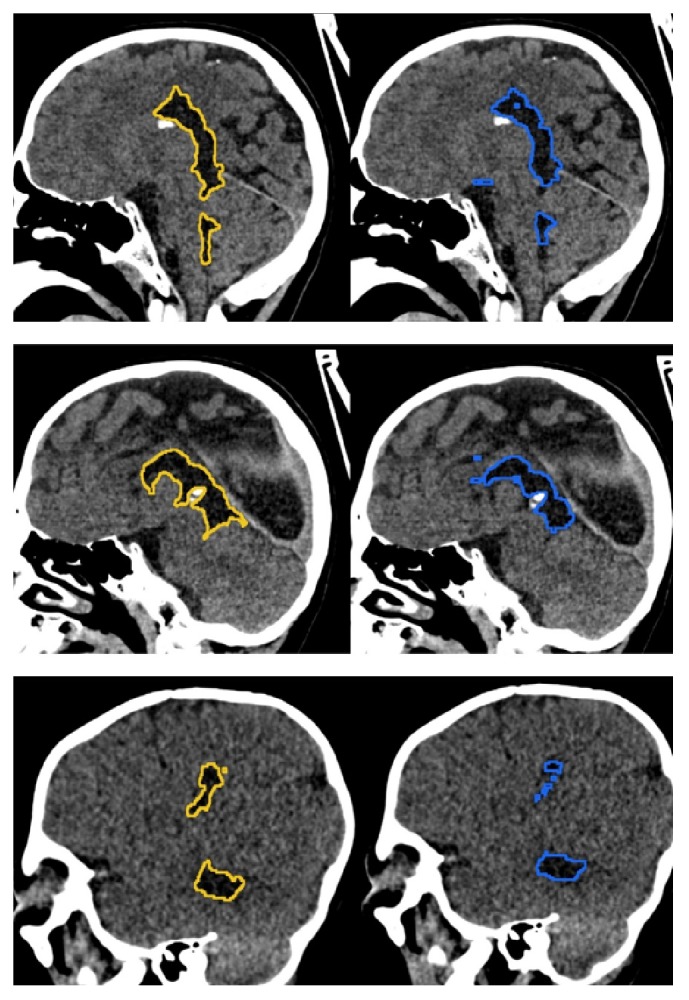
Hydrocephalus is a common neurological condition that can have traumatic ramifications and can be lethal without treatment. Nowadays, during therapy radiologists have to spend a vast amount of time assessing the volume of cerebrospinal fluid (CSF) by manual segmentation on Computed Tomography (CT) images. Further, some of the segmentations are prone to radiologist bias and high intraobserver variability. To improve this, researchers are exploring methods to automate the process, which would enable faster and more unbiased results. In this study, we propose the application of U-Net convolutional neural network in order to automatically segment CT brain scans for location of CSF. U-Net is a neural network that has proven to be successful for various interdisciplinary segmentation tasks. We optimised training using state of the art methods, including "1cycle" learning rate policy, transfer learning, generalized dice loss function, mixed float precision, self-attention, and data augmentation. Even though the study was performed using a limited amount of data (80 CT images), our experiment has shown near human-level performance. We managed to achieve a 0.917 mean dice score with 0.0352 standard deviation on cross validation across the training data and a 0.9506 mean dice score on a separate test set. To our knowledge, these results are better than any known method for CSF segmentation in hydrocephalic patients, and thus, it is promising for potential practical applications.

摘要

脑积水是一种常见的神经疾病,如果不治疗,可能会造成严重后果甚至致命。目前,在治疗过程中,放射科医生需要花费大量时间通过手动分割计算断层扫描(CT)图像中的脑脊液(CSF)体积。此外,一些分割结果容易受到放射科医生的主观偏见和观察者内变异性的影响。为了改进这一点,研究人员正在探索自动化处理的方法,这将使结果更快、更客观。在这项研究中,我们提出了应用 U-Net 卷积神经网络自动分割 CT 脑部扫描以定位 CSF。U-Net 是一种已经在各种跨学科分割任务中被证明是成功的神经网络。我们使用最先进的方法进行了训练优化,包括“1 周期”学习率策略、迁移学习、广义骰子损失函数、混合浮点精度、自注意力和数据增强。尽管该研究是使用有限数量的数据(80 个 CT 图像)进行的,但我们的实验表明,它的性能接近人类水平。我们在交叉验证中实现了 0.917 的平均骰子分数,标准偏差为 0.0352,在独立的测试集上实现了 0.9506 的平均骰子分数。据我们所知,这些结果优于任何已知的脑积水患者 CSF 分割方法,因此,它有望在实际应用中得到应用。

https://cdn.ncbi.nlm.nih.gov/pmc/blobs/0247/6642766/f2fb2ab61614/BMRI2019-3059170.005.jpg
https://cdn.ncbi.nlm.nih.gov/pmc/blobs/0247/6642766/092526875c48/BMRI2019-3059170.001.jpg
https://cdn.ncbi.nlm.nih.gov/pmc/blobs/0247/6642766/022e5c2366d5/BMRI2019-3059170.002.jpg
https://cdn.ncbi.nlm.nih.gov/pmc/blobs/0247/6642766/16cd896d26dc/BMRI2019-3059170.003.jpg
https://cdn.ncbi.nlm.nih.gov/pmc/blobs/0247/6642766/f0ca4491926e/BMRI2019-3059170.004.jpg
https://cdn.ncbi.nlm.nih.gov/pmc/blobs/0247/6642766/f2fb2ab61614/BMRI2019-3059170.005.jpg

相似文献

1
Automated Ventricular System Segmentation in Paediatric Patients Treated for Hydrocephalus Using Deep Learning Methods.基于深度学习方法对脑积水患儿进行自动心室系统分割。
Biomed Res Int. 2019 Jul 7;2019:3059170. doi: 10.1155/2019/3059170. eCollection 2019.
2
Artificial intelligence for automatic cerebral ventricle segmentation and volume calculation: a clinical tool for the evaluation of pediatric hydrocephalus.人工智能在自动脑室内层分割和容量计算中的应用:小儿脑积水评估的临床工具。
J Neurosurg Pediatr. 2020 Dec 1;27(2):131-138. doi: 10.3171/2020.6.PEDS20251. Print 2021 Feb 1.
3
Semantic segmentation of cerebrospinal fluid and brain volume with a convolutional neural network in pediatric hydrocephalus-transfer learning from existing algorithms.基于卷积神经网络的小儿脑积水脑积水分割及脑容量计算——从现有算法进行迁移学习。
Acta Neurochir (Wien). 2020 Oct;162(10):2463-2474. doi: 10.1007/s00701-020-04447-x. Epub 2020 Jun 25.
4
Fully automated intracranial ventricle segmentation on CT with 2D regional convolutional neural network to estimate ventricular volume.基于二维区域卷积神经网络的 CT 全自动颅内脑室分割以估算脑室容积。
Int J Comput Assist Radiol Surg. 2019 Nov;14(11):1923-1932. doi: 10.1007/s11548-019-02038-5. Epub 2019 Jul 26.
5
Using deep learning convolutional neural networks to automatically perform cerebral aqueduct CSF flow analysis.利用深度学习卷积神经网络自动进行脑脊髓液流动分析。
J Clin Neurosci. 2021 Aug;90:60-67. doi: 10.1016/j.jocn.2021.05.010. Epub 2021 May 24.
6
AnatomyNet: Deep learning for fast and fully automated whole-volume segmentation of head and neck anatomy.AnatomyNet:用于快速和全自动对头颈部解剖结构进行整体体积分割的深度学习方法。
Med Phys. 2019 Feb;46(2):576-589. doi: 10.1002/mp.13300. Epub 2018 Dec 17.
7
Automated Lateral Ventricular and Cranial Vault Volume Measurements in 13,851 Patients Using Deep Learning Algorithms.使用深度学习算法对13851例患者进行侧脑室和颅腔容积的自动测量
World Neurosurg. 2021 Apr;148:e363-e373. doi: 10.1016/j.wneu.2020.12.148. Epub 2021 Jan 6.
8
Data augmentation using generative adversarial networks (CycleGAN) to improve generalizability in CT segmentation tasks.使用生成对抗网络(CycleGAN)进行数据增强以提高 CT 分割任务的泛化能力。
Sci Rep. 2019 Nov 15;9(1):16884. doi: 10.1038/s41598-019-52737-x.
9
Deep learning-based segmentation of multisite disease in ovarian cancer.基于深度学习的卵巢癌多部位病变分割。
Eur Radiol Exp. 2023 Dec 7;7(1):77. doi: 10.1186/s41747-023-00388-z.
10
Deep learning for segmentation of 49 selected bones in CT scans: First step in automated PET/CT-based 3D quantification of skeletal metastases.深度学习在 CT 扫描中对 49 块选定骨骼的分割:基于 PET/CT 的骨骼转移全自动 3D 定量的第一步。
Eur J Radiol. 2019 Apr;113:89-95. doi: 10.1016/j.ejrad.2019.01.028. Epub 2019 Feb 1.

引用本文的文献

1
Deep learning for hydrocephalus prognosis: Advances, challenges, and future directions: A review.脑积水预后的深度学习:进展、挑战与未来方向:综述
Medicine (Baltimore). 2025 Jun 27;104(26):e43082. doi: 10.1097/MD.0000000000043082.
2
Volumetric measurement of cranial cavity and cerebral ventricular system with 3D Slicer software based on CT data.基于CT数据,使用3D Slicer软件对颅腔和脑室系统进行容积测量。
BMC Med Imaging. 2025 Feb 26;25(1):64. doi: 10.1186/s12880-025-01591-7.
3
Automated ventricular segmentation and shunt failure detection using convolutional neural networks.

本文引用的文献

1
Automatic Coronary Wall and Atherosclerotic Plaque Segmentation from 3D Coronary CT Angiography.从 3D 冠状动脉 CT 血管造影自动分割冠状动脉壁和动脉粥样硬化斑块。
Sci Rep. 2019 Jan 10;9(1):47. doi: 10.1038/s41598-018-37168-4.
2
Automatic Segmentation of Neonatal Ventricles from Cranial Ultrasound for Prediction of Intraventricular Hemorrhage Outcome.通过颅脑超声自动分割新生儿脑室以预测脑室内出血结局
Annu Int Conf IEEE Eng Med Biol Soc. 2018 Jul;2018:3136-3139. doi: 10.1109/EMBC.2018.8513097.
3
DRINet for Medical Image Segmentation.
使用卷积神经网络进行自动心室分割和分流失败检测。
Sci Rep. 2024 Sep 27;14(1):22166. doi: 10.1038/s41598-024-73167-4.
4
Automated segmentation of ventricular volumes and subarachnoid hemorrhage from computed tomography images: Evaluation of a rule-based pipeline approach.基于计算机断层扫描图像的心室容积和蛛网膜下腔出血自动分割:基于规则的流水线方法评估
Neuroradiol J. 2025 Feb;38(1):30-43. doi: 10.1177/19714009241260791. Epub 2024 Jun 13.
5
Current Perspectives of Artificial Intelligence in Pediatric Neuroradiology: An Overview.人工智能在儿科神经放射学中的当前视角:概述
Front Radiol. 2021 Sep 7;1:713681. doi: 10.3389/fradi.2021.713681. eCollection 2021.
6
Validation of a deep learning model for traumatic brain injury detection and NIRIS grading on non-contrast CT: a multi-reader study with promising results and opportunities for improvement.深度学习模型在非对比 CT 上检测创伤性脑损伤和 NIRIS 分级的验证:一项多读者研究,结果有前景,有改进的机会。
Neuroradiology. 2023 Nov;65(11):1605-1617. doi: 10.1007/s00234-023-03170-5. Epub 2023 Jun 3.
7
Prediction of shunt failure facilitated by rapid and accurate volumetric analysis: a single institution's preliminary experience.快速准确的容量分析有助于预测分流失败:单中心初步经验。
Childs Nerv Syst. 2022 Oct;38(10):1907-1912. doi: 10.1007/s00381-022-05552-1. Epub 2022 May 20.
8
Hydrocephalus: Ventricular Volume Quantification Using Three-Dimensional Brain CT Data and Semiautomatic Three-Dimensional Threshold-Based Segmentation Approach.脑积水:基于三维脑 CT 数据和半自动三维阈值分割方法的脑室容积量化。
Korean J Radiol. 2021 Mar;22(3):435-441. doi: 10.3348/kjr.2020.0671. Epub 2020 Oct 30.
9
Artificial Intelligence in Neuroradiology: Current Status and Future Directions.人工智能在神经放射学中的应用:现状与未来方向。
AJNR Am J Neuroradiol. 2020 Aug;41(8):E52-E59. doi: 10.3174/ajnr.A6681. Epub 2020 Jul 30.
10
Semantic segmentation of cerebrospinal fluid and brain volume with a convolutional neural network in pediatric hydrocephalus-transfer learning from existing algorithms.基于卷积神经网络的小儿脑积水脑积水分割及脑容量计算——从现有算法进行迁移学习。
Acta Neurochir (Wien). 2020 Oct;162(10):2463-2474. doi: 10.1007/s00701-020-04447-x. Epub 2020 Jun 25.
DRINet 用于医学图像分割。
IEEE Trans Med Imaging. 2018 Nov;37(11):2453-2462. doi: 10.1109/TMI.2018.2835303. Epub 2018 May 10.
4
Learning Based Segmentation of CT Brain Images: Application to Postoperative Hydrocephalic Scans.基于学习的 CT 脑图像分割:在术后脑积水扫描中的应用。
IEEE Trans Biomed Eng. 2018 Aug;65(8):1871-1884. doi: 10.1109/TBME.2017.2783305. Epub 2017 Dec 13.
5
Recent Advancements in Retinal Vessel Segmentation.视网膜血管分割的最新进展
J Med Syst. 2017 Apr;41(4):70. doi: 10.1007/s10916-017-0719-2. Epub 2017 Mar 11.
6
A combined deep-learning and deformable-model approach to fully automatic segmentation of the left ventricle in cardiac MRI.一种联合深度学习和可变形模型的方法,用于心脏 MRI 中左心室的全自动分割。
Med Image Anal. 2016 May;30:108-119. doi: 10.1016/j.media.2016.01.005. Epub 2016 Feb 6.
7
Methods on Skull Stripping of MRI Head Scan Images-a Review.磁共振成像头部扫描图像的颅骨剥离方法——综述
J Digit Imaging. 2016 Jun;29(3):365-79. doi: 10.1007/s10278-015-9847-8.
8
Pediatric hydrocephalus: systematic literature review and evidence-based guidelines. Part 1: Introduction and methodology.小儿脑积水:系统文献综述与循证指南。第1部分:引言与方法
J Neurosurg Pediatr. 2014 Nov;14 Suppl 1:3-7. doi: 10.3171/2014.7.PEDS14321.
9
Volumetric brain analysis in neurosurgery: Part 1. Particle filter segmentation of brain and cerebrospinal fluid growth dynamics from MRI and CT images.神经外科中的脑容量分析:第1部分。基于MRI和CT图像的脑和脑脊液生长动力学的粒子滤波分割
J Neurosurg Pediatr. 2015 Feb;15(2):113-24. doi: 10.3171/2014.9.PEDS12426. Epub 2014 Nov 28.
10
Automatic whole brain MRI segmentation of the developing neonatal brain.自动全脑 MRI 新生儿脑发育的分段。
IEEE Trans Med Imaging. 2014 Sep;33(9):1818-31. doi: 10.1109/TMI.2014.2322280. Epub 2014 May 6.