1Department of Neurosurgery, Stanford University School of Medicine.
2Stanford University School of Medicine.
J Neurosurg Pediatr. 2020 Dec 1;27(2):131-138. doi: 10.3171/2020.6.PEDS20251. Print 2021 Feb 1.
Imaging evaluation of the cerebral ventricles is important for clinical decision-making in pediatric hydrocephalus. Although quantitative measurements of ventricular size, over time, can facilitate objective comparison, automated tools for calculating ventricular volume are not structured for clinical use. The authors aimed to develop a fully automated deep learning (DL) model for pediatric cerebral ventricle segmentation and volume calculation for widespread clinical implementation across multiple hospitals.
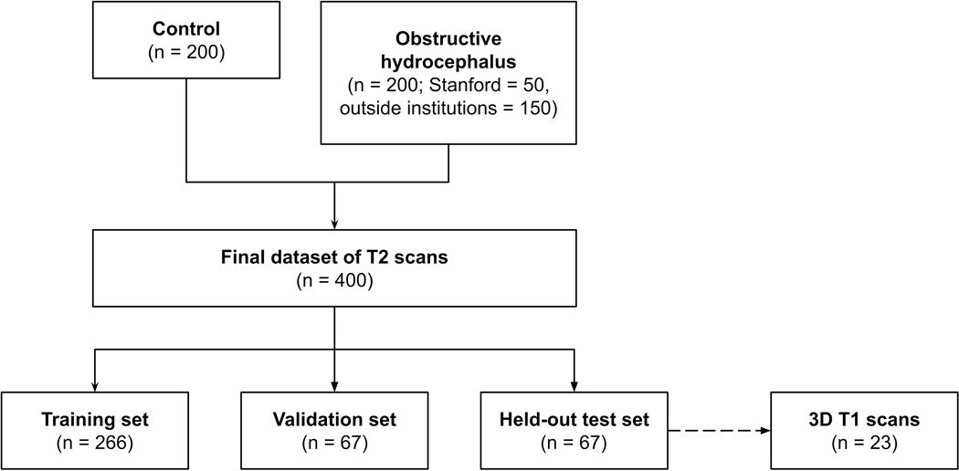
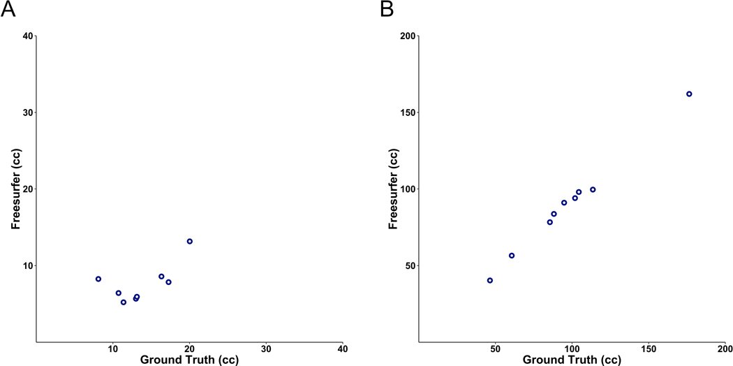
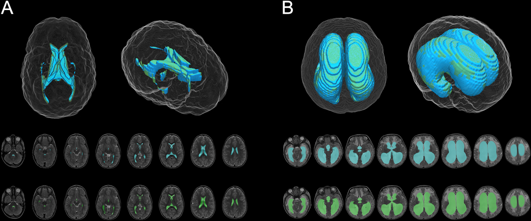
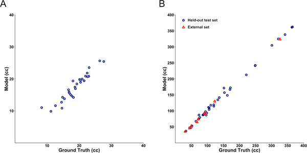
The study cohort consisted of 200 children with obstructive hydrocephalus from four pediatric hospitals, along with 199 controls. Manual ventricle segmentation and volume calculation values served as "ground truth" data. An encoder-decoder convolutional neural network architecture, in which T2-weighted MR images were used as input, automatically delineated the ventricles and output volumetric measurements. On a held-out test set, segmentation accuracy was assessed using the Dice similarity coefficient (0 to 1) and volume calculation was assessed using linear regression. Model generalizability was evaluated on an external MRI data set from a fifth hospital. The DL model performance was compared against FreeSurfer research segmentation software.
Model segmentation performed with an overall Dice score of 0.901 (0.946 in hydrocephalus, 0.856 in controls). The model generalized to external MR images from a fifth pediatric hospital with a Dice score of 0.926. The model was more accurate than FreeSurfer, with faster operating times (1.48 seconds per scan).
The authors present a DL model for automatic ventricle segmentation and volume calculation that is more accurate and rapid than currently available methods. With near-immediate volumetric output and reliable performance across institutional scanner types, this model can be adapted to the real-time clinical evaluation of hydrocephalus and improve clinician workflow.
脑室内腔的影像学评估对小儿脑积水的临床决策至关重要。尽管脑室大小的定量测量可以方便客观比较,但用于计算脑室容积的自动化工具尚未针对临床使用进行构建。作者旨在开发一种全自动深度学习(DL)模型,用于小儿脑室内腔分割和容积计算,以便在多家医院广泛临床应用。
研究队列包括来自 4 家儿童医院的 200 名梗阻性脑积水患儿和 199 名对照患儿。手动脑室分割和容积计算值作为“金标准”数据。采用编码器-解码器卷积神经网络架构,以 T2 加权磁共振图像作为输入,自动勾画脑室并输出容积测量值。在保留的测试集中,使用 Dice 相似系数(0 到 1)评估分割准确性,使用线性回归评估容积计算准确性。在来自第 5 家医院的外部 MRI 数据集上评估模型的泛化能力。将 DL 模型的性能与 FreeSurfer 研究分割软件进行比较。
模型分割的总体 Dice 得分为 0.901(脑积水为 0.946,对照组为 0.856)。该模型可推广至来自第 5 家儿童医院的外部 MRI 图像,Dice 得分为 0.926。该模型比 FreeSurfer 更准确,且操作时间更快(每次扫描 1.48 秒)。
作者提出了一种用于自动脑室分割和容积计算的 DL 模型,其比现有的方法更准确、快速。该模型具有接近即时的容积输出和可靠的性能,适用于不同机构扫描仪类型,可以适应脑积水的实时临床评估,并改善临床医生的工作流程。