Department of Respiratory Care and Sleep Control Medicine, Kyoto University, Kyoto, Japan.
Department of Neurology, Kyoto University, Kyoto, Japan.
Brain Behav. 2019 Sep;9(9):e01366. doi: 10.1002/brb3.1366. Epub 2019 Jul 30.
We aimed at clarifying the clinical significance of the responses evoked by human entorhinal cortex (EC) electrical stimulation by means of cortico-cortical evoked potentials (CCEPs).
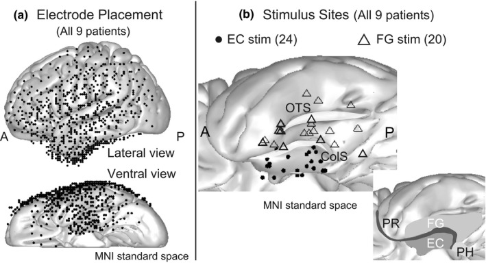
We enrolled nine patients with medically intractable medial temporal lobe epilepsy who underwent invasive presurgical evaluations with subdural or depth electrodes. Single-pulse electrical stimulation was delivered to the EC and fusiform gyrus (FG), and their evoked potentials were compared. The correlation between the evoked potentials and Wechsler Memory Scale-Revised (WMS-R) score was analyzed to investigate whether memory circuit was involved in the generation of the evoked potentials.
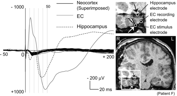
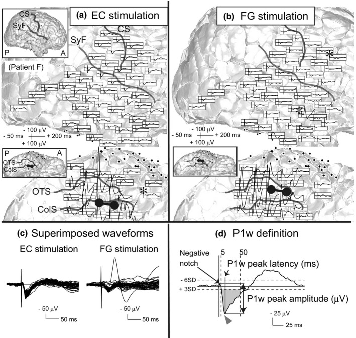
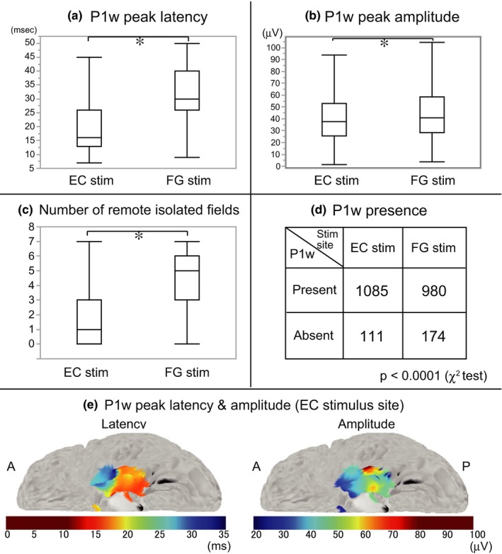
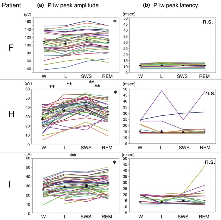
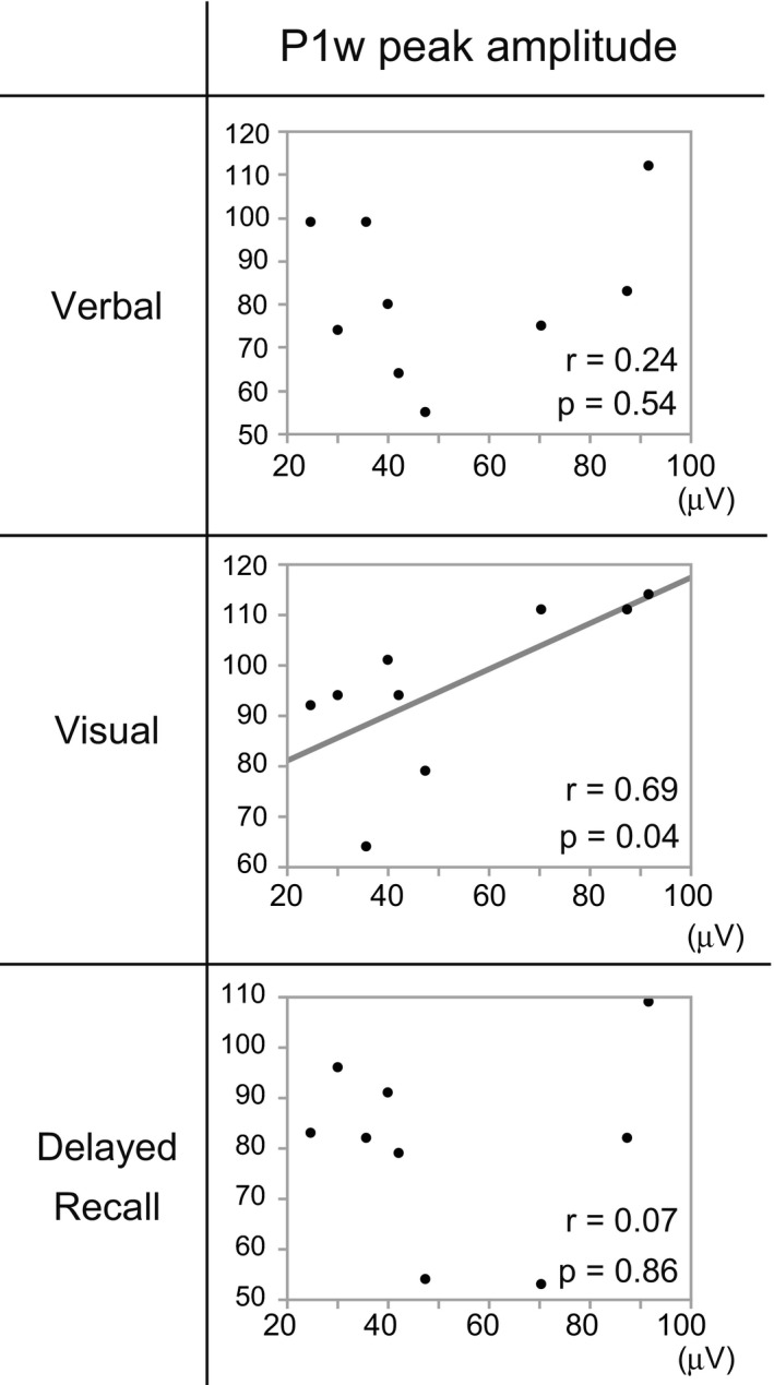
In most electrodes placed on the neocortex, EC stimulation induced unique evoked potentials with positive polarity, termed as "widespread P1" (P1w). Compared with FG stimulation, P1w induced by EC stimulation were distinguished by their high occurrence rate, short peak latency (mean: 20.1 ms), small peak amplitude, and waveform uniformity among different recording sites. A stimulation of more posterior parts of the EC induced P1w with shorter latency and larger amplitude. P1w peak amplitude had a positive correlation (r = .69) with the visual memory score of the WMS-R. In one patient, with depth electrode implanted into the hippocampus, the giant evoked potentials were recorded in the electrodes of the anterior hippocampus and EC near the stimulus site.
The human EC electrical stimulation evoked the short-latency potentials in the broad neocortical regions. The origin of P1w remains unclear, although the limited evidence suggests that P1w is the far-field potential by the volume conduction of giant evoked potential from the EC itself and hippocampus. The significance of the present study is that those evoked potentials may be a potential biomarker of memory impairment in various neurological diseases, and we provided direct evidence for the functional subdivisions along the anterior-posterior axis in the human EC.
通过皮质-皮质诱发电位(CCEPs)阐明人类内嗅皮层(EC)电刺激引起的反应的临床意义。
我们纳入了 9 名接受过有创术前评估的药物难治性内侧颞叶癫痫患者,这些患者使用了硬膜下或深部电极。对 EC 和梭状回(FG)进行单脉冲电刺激,并比较它们的诱发电位。分析诱发电位与韦氏记忆量表修订版(WMS-R)评分之间的相关性,以研究记忆回路是否参与了诱发电位的产生。
在放置在新皮层上的大多数电极上,EC 刺激诱导出具有正极性的独特诱发电位,称为“广泛 P1”(P1w)。与 FG 刺激相比,EC 刺激诱导的 P1w 的特征在于其高发生率、短潜伏期(平均 20.1ms)、小峰值幅度和不同记录部位之间的波形一致性。EC 的更后部分的刺激诱导出潜伏期更短、幅度更大的 P1w。P1w 峰值幅度与 WMS-R 的视觉记忆评分呈正相关(r=.69)。在一名患者中,深部电极植入海马中,在刺激部位附近的电极中记录到了前海马和 EC 的巨大诱发电位。
人类 EC 电刺激在广泛的新皮层区域引起短潜伏期电位。尽管有限的证据表明 P1w 是来自 EC 本身和海马的巨大诱发电位的容积传导的远场电位,但 P1w 的起源尚不清楚。本研究的意义在于,这些诱发电位可能是各种神经疾病中记忆障碍的潜在生物标志物,并且我们为人类 EC 沿前后轴的功能分区提供了直接证据。