• 文献检索
  • 文档翻译
  • 深度研究
  • 学术资讯
  • Suppr Zotero 插件Zotero 插件
  • 邀请有礼
  • 套餐&价格
  • 历史记录
应用&插件
Suppr Zotero 插件Zotero 插件浏览器插件Mac 客户端Windows 客户端微信小程序
定价
高级版会员购买积分包购买API积分包
服务
文献检索文档翻译深度研究API 文档MCP 服务
关于我们
关于 Suppr公司介绍联系我们用户协议隐私条款
关注我们

Suppr 超能文献

核心技术专利:CN118964589B侵权必究
粤ICP备2023148730 号-1Suppr @ 2026

文献检索

告别复杂PubMed语法,用中文像聊天一样搜索,搜遍4000万医学文献。AI智能推荐,让科研检索更轻松。

立即免费搜索

文件翻译

保留排版,准确专业,支持PDF/Word/PPT等文件格式,支持 12+语言互译。

免费翻译文档

深度研究

AI帮你快速写综述,25分钟生成高质量综述,智能提取关键信息,辅助科研写作。

立即免费体验

比较突触前和突触后线粒体的三维超微结构。

Comparing 3D ultrastructure of presynaptic and postsynaptic mitochondria.

作者信息

Delgado Thomas, Petralia Ronald S, Freeman David W, Sedlacek Miloslav, Wang Ya-Xian, Brenowitz Stephan D, Sheu Shu-Hsien, Gu Jeffrey W, Kapogiannis Dimitrios, Mattson Mark P, Yao Pamela J

机构信息

Laboratory of Neurosciences, NIA/NIH, Baltimore, Maryland 21224, USA.

Advanced Imaging Core, NIDCD/NIH, Bethesda, Maryland 20892, USA.

出版信息

Biol Open. 2019 Aug 1;8(8):bio044834. doi: 10.1242/bio.044834.

DOI:10.1242/bio.044834
PMID:31362947
原文链接:https://pmc.ncbi.nlm.nih.gov/articles/PMC6737966/
Abstract

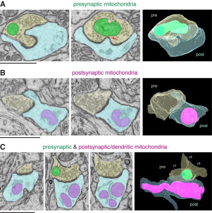
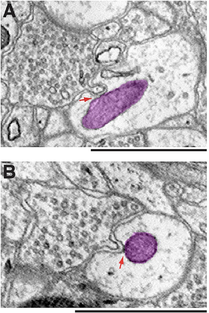
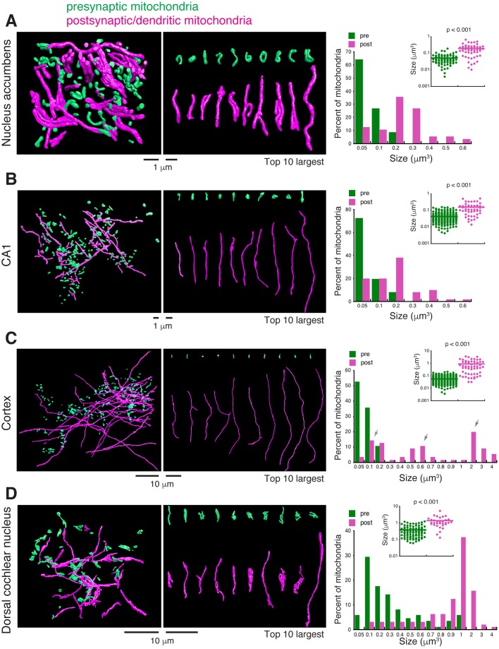
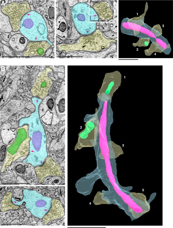
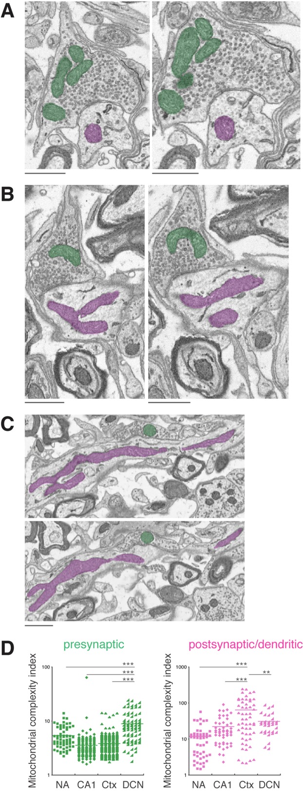
Serial-section electron microscopy such as FIB-SEM (focused ion beam scanning electron microscopy) has become an important tool for neuroscientists to trace the trajectories and global architecture of neural circuits in the brain, as well as to visualize the 3D ultrastructure of cellular organelles in neurons. In this study, we examined 3D features of mitochondria in electron microscope images generated from serial sections of four regions of mouse brains: nucleus accumbens (NA), hippocampal CA1, somatosensory cortex and dorsal cochlear nucleus (DCN). We compared mitochondria in the presynaptic terminals to those in the postsynaptic/dendritic compartments, and we focused on the shape and size of mitochondria. A common feature of mitochondria among the four brain regions is that presynaptic mitochondria generally are small and short, and most of them do not extend beyond presynaptic terminals. In contrast, the majority of postsynaptic/dendritic mitochondria are large and many of them spread through significant portions of the dendrites. Comparing among the brain areas, the cerebral cortex and DCN have even larger postsynaptic/dendritic mitochondria than the NA and CA1. Our analysis reveals that mitochondria in neurons are differentially sized and arranged according to their subcellular locations, suggesting a spatial organizing principle of mitochondria at the synapse.

摘要

诸如聚焦离子束扫描电子显微镜(FIB-SEM)之类的连续切片电子显微镜已成为神经科学家追踪大脑中神经回路轨迹和整体结构,以及可视化神经元细胞器三维超微结构的重要工具。在本研究中,我们检查了从小鼠大脑四个区域(伏隔核(NA)、海马CA1、躯体感觉皮层和背侧耳蜗核(DCN))的连续切片生成的电子显微镜图像中线粒体的三维特征。我们比较了突触前终末中的线粒体与突触后/树突区室中的线粒体,并着重研究了线粒体的形状和大小。四个脑区线粒体的一个共同特征是,突触前线粒体通常较小且较短,并且大多数不会延伸到突触前终末之外。相比之下,大多数突触后/树突线粒体较大,并且其中许多分布在树突的大部分区域。在脑区之间进行比较时,大脑皮层和DCN的突触后/树突线粒体比NA和CA1的更大。我们的分析表明,神经元中的线粒体根据其亚细胞位置大小不同且排列方式各异,这表明突触处线粒体存在空间组织原则。

https://cdn.ncbi.nlm.nih.gov/pmc/blobs/3b04/6737966/2923db5146f2/biolopen-8-044834-g5.jpg
https://cdn.ncbi.nlm.nih.gov/pmc/blobs/3b04/6737966/0874f84cd0f8/biolopen-8-044834-g1.jpg
https://cdn.ncbi.nlm.nih.gov/pmc/blobs/3b04/6737966/fc423a88b268/biolopen-8-044834-g2.jpg
https://cdn.ncbi.nlm.nih.gov/pmc/blobs/3b04/6737966/ed928025a810/biolopen-8-044834-g3.jpg
https://cdn.ncbi.nlm.nih.gov/pmc/blobs/3b04/6737966/c076df83bc22/biolopen-8-044834-g4.jpg
https://cdn.ncbi.nlm.nih.gov/pmc/blobs/3b04/6737966/2923db5146f2/biolopen-8-044834-g5.jpg

相似文献

1
Comparing 3D ultrastructure of presynaptic and postsynaptic mitochondria.比较突触前和突触后线粒体的三维超微结构。
Biol Open. 2019 Aug 1;8(8):bio044834. doi: 10.1242/bio.044834.
2
FIB/SEM technology and high-throughput 3D reconstruction of dendritic spines and synapses in GFP-labeled adult-generated neurons.FIB/SEM技术与绿色荧光蛋白标记的成年新生神经元中树突棘和突触的高通量三维重建
Front Neuroanat. 2015 May 21;9:60. doi: 10.3389/fnana.2015.00060. eCollection 2015.
3
Geometry and the Organizational Principle of Spine Synapses along a Dendrite.几何与沿着树突的脊柱突触的组织原则。
eNeuro. 2020 Oct 27;7(6). doi: 10.1523/ENEURO.0248-20.2020. Print 2020 Nov-Dec.
4
Unique synaptic topography of crest-type synapses in the interpeduncular nucleus.中脑脚间核中丘型突触的独特突触拓扑结构。
Biochem Biophys Res Commun. 2020 Sep 10;530(1):130-135. doi: 10.1016/j.bbrc.2020.06.046. Epub 2020 Jul 30.
5
Volumetric 3D reconstruction of plant leaf cells using SEM, ion milling, TEM, and serial sectioning.使用 SEM、离子铣削、TEM 和连续切片对植物叶片细胞进行体积 3D 重建。
Planta. 2022 May 6;255(6):118. doi: 10.1007/s00425-022-03905-3.
6
3D Reconstruction and Analysis of Thin Subcellular Neuronal Structures using Focused-Ion Beam Scanning Electron Microscopy Data.使用聚焦离子束扫描电子显微镜数据进行薄细胞神经结构的 3D 重建和分析。
J Vis Exp. 2021 Sep 20(175). doi: 10.3791/63030.
7
Synaptic spinules are reliable indicators of excitatory presynaptic bouton size and strength and are ubiquitous components of excitatory synapses in CA1 hippocampus.突触棘是兴奋性突触前终扣大小和强度的可靠指标,是海马体CA1区兴奋性突触的普遍组成部分。
Front Synaptic Neurosci. 2022 Aug 11;14:968404. doi: 10.3389/fnsyn.2022.968404. eCollection 2022.
8
Beyond counts and shapes: studying pathology of dendritic spines in the context of the surrounding neuropil through serial section electron microscopy.超越数量和形态:通过连续切片电子显微镜研究树突棘在周围神经间质中的病理。
Neuroscience. 2013 Oct 22;251:75-89. doi: 10.1016/j.neuroscience.2012.04.061. Epub 2012 May 1.
9
Occurrence and three-dimensional structure of multiple synapses between individual radiatum axons and their target pyramidal cells in hippocampal area CA1.海马体CA1区单个辐射层轴突与其目标锥体细胞之间多个突触的发生及三维结构。
J Neurosci. 1993 Sep;13(9):3736-48. doi: 10.1523/JNEUROSCI.13-09-03736.1993.
10
Mapping and morphometric analysis of synapses and spines on fusiform cells in the dorsal cochlear nucleus.对耳蜗背核中梭形细胞上突触和棘的映射和形态计量分析。
Front Syst Neurosci. 2014 Sep 23;8:167. doi: 10.3389/fnsys.2014.00167. eCollection 2014.

引用本文的文献

1
MCU expression in hippocampal CA2 neurons modulates dendritic mitochondrial morphology and synaptic plasticity.海马体CA2神经元中的MCU表达调节树突线粒体形态和突触可塑性。
Sci Rep. 2025 Feb 6;15(1):4540. doi: 10.1038/s41598-025-85958-4.
2
Memory engram synapse 3D molecular architecture visualized by cryoCLEM-guided cryoET.通过冷冻CLEM引导的冷冻电镜断层扫描可视化的记忆印迹突触三维分子结构。
bioRxiv. 2025 Jan 12:2025.01.09.632151. doi: 10.1101/2025.01.09.632151.
3
Ultrastructural Analysis Reveals Mitochondrial Placement Independent of Synapse Placement in Fine Caliber C. elegans Neurons.

本文引用的文献

1
Mitochondria in hippocampal presynaptic and postsynaptic compartments differ in size as well as intensity.海马体突触前和突触后区室中的线粒体在大小和强度上存在差异。
Matters (Zur). 2017;2017. doi: 10.19185/matters.201711000009. Epub 2017 Nov 30.
2
Quantitative 3D Mapping of the Human Skeletal Muscle Mitochondrial Network.人体骨骼肌线粒体网络的定量三维映射
Cell Rep. 2019 Apr 2;27(1):321. doi: 10.1016/j.celrep.2019.03.051.
3
Cortical column and whole-brain imaging with molecular contrast and nanoscale resolution.皮层柱和全脑成像的分子对比和纳米级分辨率。
超微结构分析揭示了秀丽隐杆线虫细口径神经元中线粒体的定位独立于突触定位。
J Comp Neurol. 2024 Dec;532(12):e70002. doi: 10.1002/cne.70002.
4
Mitochondrial Dynamics and mRNA Translation: A Local Synaptic Tale.线粒体动力学与mRNA翻译:一个局部突触的故事。
Biology (Basel). 2024 Sep 23;13(9):746. doi: 10.3390/biology13090746.
5
Mitochondrial plasticity and synaptic plasticity crosstalk; in health and Alzheimer's disease.线粒体可塑性和突触可塑性的相互作用;在健康和阿尔茨海默病中的作用。
CNS Neurosci Ther. 2024 Aug;30(8):e14897. doi: 10.1111/cns.14897.
6
Postsynaptic mitochondria are positioned to support functional diversity of dendritic spines.突触后线粒体的定位支持树突棘功能的多样性。
Elife. 2023 Dec 7;12:RP89682. doi: 10.7554/eLife.89682.
7
MCU-enriched dendritic mitochondria regulate plasticity in distinct hippocampal circuits.富含MCU的树突状线粒体调节不同海马回路中的可塑性。
bioRxiv. 2024 Apr 3:2023.11.10.566606. doi: 10.1101/2023.11.10.566606.
8
Axonal energy metabolism, and the effects in aging and neurodegenerative diseases.轴突能量代谢,以及在衰老和神经退行性疾病中的作用。
Mol Neurodegener. 2023 Jul 20;18(1):49. doi: 10.1186/s13024-023-00634-3.
9
Instance segmentation of mitochondria in electron microscopy images with a generalist deep learning model trained on a diverse dataset.使用在多样化数据集上训练的通用深度学习模型对电子显微镜图像中的线粒体进行实例分割。
Cell Syst. 2023 Jan 18;14(1):58-71.e5. doi: 10.1016/j.cels.2022.12.006.
10
A serotonergic axon-cilium synapse drives nuclear signaling to alter chromatin accessibility.一种血清素能轴突-纤毛突触驱动核信号转导改变染色质可及性。
Cell. 2022 Sep 1;185(18):3390-3407.e18. doi: 10.1016/j.cell.2022.07.026.
Science. 2019 Jan 18;363(6424). doi: 10.1126/science.aau8302.
4
Spatially Stable Mitochondrial Compartments Fuel Local Translation during Plasticity.空间稳定的线粒体隔室为可塑性期间的局部翻译提供燃料。
Cell. 2019 Jan 10;176(1-2):73-84.e15. doi: 10.1016/j.cell.2018.12.013. Epub 2019 Jan 3.
5
MFF-dependent mitochondrial fission regulates presynaptic release and axon branching by limiting axonal mitochondria size.MFF 依赖性线粒体裂变通过限制轴突线粒体大小来调节突触前释放和轴突分支。
Nat Commun. 2018 Nov 27;9(1):5008. doi: 10.1038/s41467-018-07416-2.
6
Visualizing Intracellular Organelle and Cytoskeletal Interactions at Nanoscale Resolution on Millisecond Timescales.在毫秒时间尺度上以纳米分辨率可视化细胞内细胞器和细胞骨架相互作用。
Cell. 2018 Nov 15;175(5):1430-1442.e17. doi: 10.1016/j.cell.2018.09.057. Epub 2018 Oct 25.
7
VAST (Volume Annotation and Segmentation Tool): Efficient Manual and Semi-Automatic Labeling of Large 3D Image Stacks.VAST(体积注释和分割工具):高效的大型 3D 图像堆栈手动和半自动标注。
Front Neural Circuits. 2018 Oct 16;12:88. doi: 10.3389/fncir.2018.00088. eCollection 2018.
8
Interacting organelles.相互作用的细胞器。
Curr Opin Cell Biol. 2018 Aug;53:84-91. doi: 10.1016/j.ceb.2018.06.003. Epub 2018 Jul 2.
9
Invaginating Structures in Mammalian Synapses.哺乳动物突触中的内陷结构。
Front Synaptic Neurosci. 2018 Apr 5;10:4. doi: 10.3389/fnsyn.2018.00004. eCollection 2018.
10
Mitochondrial Ultrastructure Is Coupled to Synaptic Performance at Axonal Release Sites.线粒体超微结构与轴突释放部位突触性能相偶联。
eNeuro. 2018 Jan 29;5(1). doi: 10.1523/ENEURO.0390-17.2018. eCollection 2018 Jan-Feb.