• 文献检索
  • 文档翻译
  • 深度研究
  • 学术资讯
  • Suppr Zotero 插件Zotero 插件
  • 邀请有礼
  • 套餐&价格
  • 历史记录
应用&插件
Suppr Zotero 插件Zotero 插件浏览器插件Mac 客户端Windows 客户端微信小程序
定价
高级版会员购买积分包购买API积分包
服务
文献检索文档翻译深度研究API 文档MCP 服务
关于我们
关于 Suppr公司介绍联系我们用户协议隐私条款
关注我们

Suppr 超能文献

核心技术专利:CN118964589B侵权必究
粤ICP备2023148730 号-1Suppr @ 2026

文献检索

告别复杂PubMed语法,用中文像聊天一样搜索,搜遍4000万医学文献。AI智能推荐,让科研检索更轻松。

立即免费搜索

文件翻译

保留排版,准确专业,支持PDF/Word/PPT等文件格式,支持 12+语言互译。

免费翻译文档

深度研究

AI帮你快速写综述,25分钟生成高质量综述,智能提取关键信息,辅助科研写作。

立即免费体验

两种类型的肱骨髁上骨折内固定在解剖模型中的生物力学分析

Biomechanical Analysis of Two Types of Humerus Supracondylar Fracture Fixation in Anatomical Model.

作者信息

Nunes Marcos Ceita, Posser Ticiano Dozza, Israel Charles Leonardo, Spinelli Leandro de Freitas, Calieron Luis Gustavo, Kim Jung Ho

机构信息

Serviço de Ortopedia Pediátrica, Hospital São Vicente de Paulo, Passo Fundo, RS, Brasil.

Laboratório de Bioengenharia, Universidade de Passo Fundo, Passo Fundo, RS, Brasil.

出版信息

Rev Bras Ortop (Sao Paulo). 2019 May;54(3):261-267. doi: 10.1055/s-0039-1688756. Epub 2019 Jun 27.

DOI:10.1055/s-0039-1688756
PMID:31363279
原文链接:https://pmc.ncbi.nlm.nih.gov/articles/PMC6597430/
Abstract

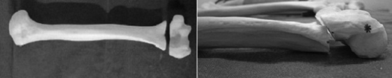
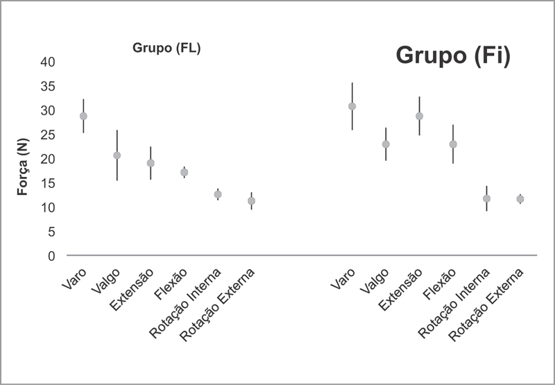
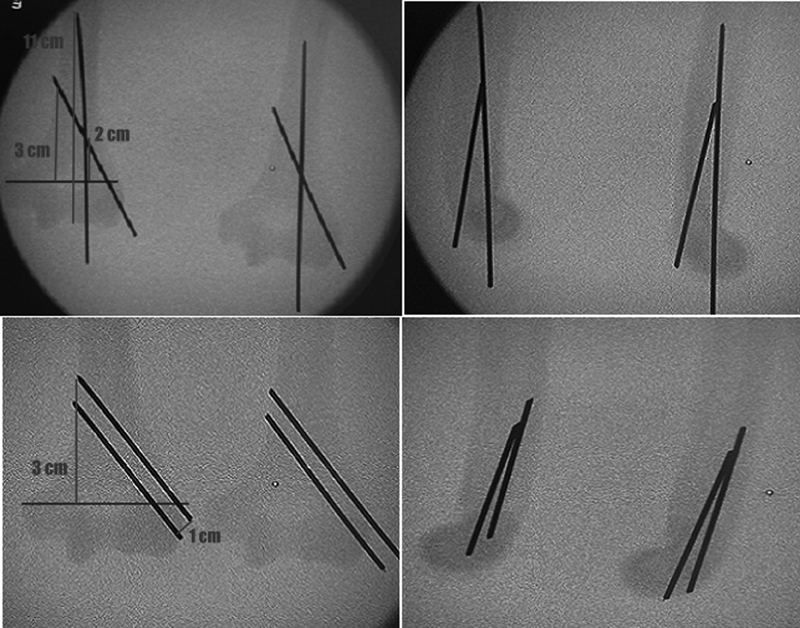
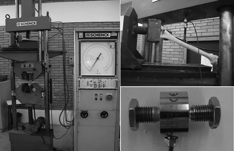
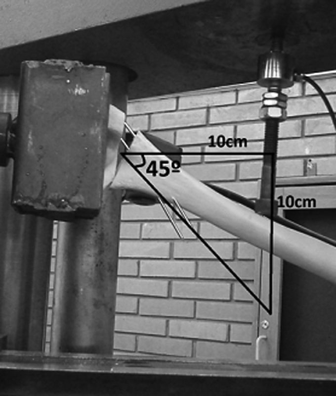
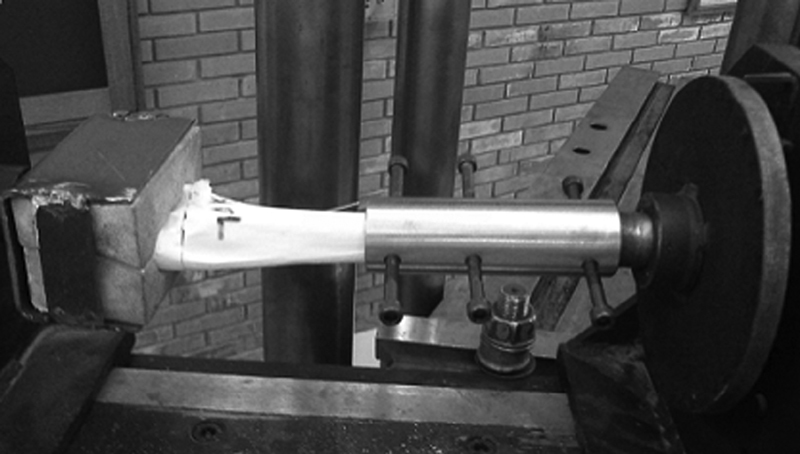
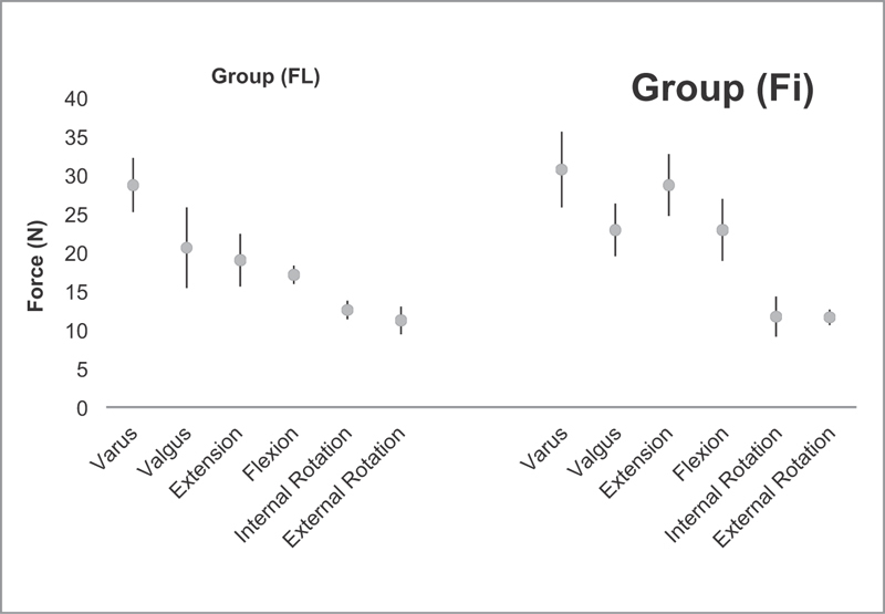
To analyze the stability of humerus supracondylar fracture fixation with Kirschner wires comparing intramedullary and lateral (Fi), and two parallel lateral wires (FL) fixation in experimental models, to define which configuration presents greater stability.  A total of 72 synthetic humeri were cross-sectioned to simulate the fracture. These bones were divided into two equal groups and the fractures were fixed with parallel Kirschner wires (FL) and with a lateral and intramedullary (Fi) wire. Then, the test specimens were subjected to stress load tests on a universal test machine, measured in Newtons (N). Each group was subdivided into varus load, valgus, extension, flexion, external rotation and internal rotation. An analysis of the data was performed comparing the subgroups of the FL group with their respective subgroups of the Fi group through the two-tailed t test.  The two-tailed t test showed that in 4 of the 6 evaluated conditions there was no significant statistical difference between the groups (  > 0.05). We have found a significant difference between the group with extension load with a mean of 19 N (FL group) and of 28.7 N (Fi group) (  = 0.004), and also between the groups with flexural load with the mean of the forces recorded in the FL group of 17.1 N and of 22.9 N in the Fi group (  = 0.01).  Fixation with one intramedullary wire and one lateral wire, considering loads in extension and flexion, presents greater stability when compared to a fixation with two lateral wires, suggesting similar clinical results.

摘要

通过比较克氏针髓内和外侧固定(Fi)以及两根平行外侧克氏针(FL)固定在实验模型中对肱骨髁上骨折固定的稳定性,以确定哪种构型具有更高的稳定性。总共72根合成肱骨被横切以模拟骨折。这些骨头被分成两组,骨折分别用平行克氏针(FL)和一根外侧及一根髓内克氏针(Fi)固定。然后,测试标本在万能试验机上进行应力加载测试,以牛顿(N)为单位测量。每组再细分为内翻负荷、外翻、伸展、屈曲、外旋和内旋。通过双尾t检验对FL组的亚组与其相应的Fi组亚组进行数据比较分析。双尾t检验表明,在6种评估条件中的4种条件下,两组之间没有显著的统计学差异(P>0.05)。我们发现在伸展负荷组中,平均力为19 N(FL组)和28.7 N(Fi组)之间存在显著差异(P = 0.004),在屈曲负荷组中,FL组记录的平均力为17.1 N,Fi组为22.9 N之间也存在显著差异(P = 0.01)。考虑到伸展和屈曲负荷,一根髓内针和一根外侧针固定比两根外侧针固定具有更高的稳定性,提示临床结果相似。

https://cdn.ncbi.nlm.nih.gov/pmc/blobs/abd8/6597430/127784fe0b2e/10-1055-s-0039-1688756-i170350pt-6.jpg
https://cdn.ncbi.nlm.nih.gov/pmc/blobs/abd8/6597430/cfff699a7b20/10-1055-s-0039-1688756-i170350en-1.jpg
https://cdn.ncbi.nlm.nih.gov/pmc/blobs/abd8/6597430/494f4dca03d8/10-1055-s-0039-1688756-i170350en-2.jpg
https://cdn.ncbi.nlm.nih.gov/pmc/blobs/abd8/6597430/551a49d343b6/10-1055-s-0039-1688756-i170350en-3.jpg
https://cdn.ncbi.nlm.nih.gov/pmc/blobs/abd8/6597430/1dcedb394ba1/10-1055-s-0039-1688756-i170350en-4.jpg
https://cdn.ncbi.nlm.nih.gov/pmc/blobs/abd8/6597430/64dec3dde929/10-1055-s-0039-1688756-i170350en-5.jpg
https://cdn.ncbi.nlm.nih.gov/pmc/blobs/abd8/6597430/395c28e86507/10-1055-s-0039-1688756-i170350en-6.jpg
https://cdn.ncbi.nlm.nih.gov/pmc/blobs/abd8/6597430/710b60d18b35/10-1055-s-0039-1688756-i170350pt-1.jpg
https://cdn.ncbi.nlm.nih.gov/pmc/blobs/abd8/6597430/a980660d84f6/10-1055-s-0039-1688756-i170350pt-2.jpg
https://cdn.ncbi.nlm.nih.gov/pmc/blobs/abd8/6597430/213380efee49/10-1055-s-0039-1688756-i170350pt-3.jpg
https://cdn.ncbi.nlm.nih.gov/pmc/blobs/abd8/6597430/15d648baae68/10-1055-s-0039-1688756-i170350pt-4.jpg
https://cdn.ncbi.nlm.nih.gov/pmc/blobs/abd8/6597430/2dddad9d715e/10-1055-s-0039-1688756-i170350pt-5.jpg
https://cdn.ncbi.nlm.nih.gov/pmc/blobs/abd8/6597430/127784fe0b2e/10-1055-s-0039-1688756-i170350pt-6.jpg

相似文献

1
Biomechanical Analysis of Two Types of Humerus Supracondylar Fracture Fixation in Anatomical Model.两种类型的肱骨髁上骨折内固定在解剖模型中的生物力学分析
Rev Bras Ortop (Sao Paulo). 2019 May;54(3):261-267. doi: 10.1055/s-0039-1688756. Epub 2019 Jun 27.
2
Biomechanical analysis between Orthofix® external fixator and different K-wire configurations for pediatric supracondylar humerus fractures.Orthofix®外固定器与不同克氏针配置用于小儿肱骨髁上骨折的生物力学分析
J Orthop Surg Res. 2018 Jul 28;13(1):188. doi: 10.1186/s13018-018-0893-z.
3
Comparison of Different Fixation Confi gurations in Different Types of High Pediatric Supracondylar Humerus Fractures: a Finite Element Analysis.不同类型儿童肱骨髁上骨折不同固定构型的比较:有限元分析。
Acta Chir Orthop Traumatol Cech. 2023;90(6):408-415.
4
Mechanical stability study of three techniques used in the fixation of transverse and oblique metaphyseal-diaphyseal junction fractures of the distal humerus in children: a finite element analysis.三种技术固定儿童肱骨远端干骺端横斜骨折的机械稳定性研究:有限元分析。
J Orthop Surg Res. 2020 Jan 31;15(1):34. doi: 10.1186/s13018-020-1564-4.
5
Biomechanical analysis of supracondylar humerus fracture pinning for slightly malreduced fractures.轻度复位不良的肱骨髁上骨折克氏针固定的生物力学分析
J Pediatr Orthop. 2008 Oct-Nov;28(7):766-72. doi: 10.1097/BPO.0b013e318186bdcd.
6
Biomechanical analysis of supracondylar humerus fracture pinning for fractures with coronal lateral obliquity.肱骨髁上骨折冠状面外侧倾斜骨折穿针固定的生物力学分析
J Pediatr Orthop. 2012 Mar;32(2):196-200. doi: 10.1097/BPO.0b013e318242a99a.
7
The effect of the angle between fracture line and Kirschner wires on stability in supracondylar humerus fractures treated with Kirschner wire fixation: A finite element analysis.骨折线与克氏针之间角度对克氏针固定治疗肱骨髁上骨折稳定性的影响:有限元分析。
Jt Dis Relat Surg. 2021;32(1):75-84. doi: 10.5606/ehc.2021.77279. Epub 2021 Jan 6.
8
[Three-dimensional finite element analysis of Kirschner wire fixation configuration for supracondylar fracture of humerus fracture in children].儿童肱骨髁上骨折克氏针内固定构型的三维有限元分析
Zhongguo Gu Shang. 2020 Oct 25;33(10):948-53. doi: 10.12200/j.issn.1003-0034.2020.10.012.
9
Biomechanical analysis of lateral pin placements for pediatric supracondylar humerus fractures.儿童肱骨髁上骨折外侧克氏针置入的生物力学分析
J Pediatr Orthop. 2010 Mar;30(2):135-9. doi: 10.1097/BPO.0b013e3181cfcd14.
10
Finite element analysis of modified Slongo's external fixation in the treatment of supracondylar humeral fractures in older children.改良Slongo外固定治疗大龄儿童肱骨髁上骨折的有限元分析
Medicine (Baltimore). 2024 May 3;103(18):e37979. doi: 10.1097/MD.0000000000037979.

引用本文的文献

1
Proximal lateral entry technique for the treatment of supracondylar humeral fractures in children and intraoperative nerve protection: a novel technique.儿童肱骨髁上骨折治疗的近端外侧入路技术及术中神经保护:一种新技术
BMC Musculoskelet Disord. 2025 Jul 5;26(1):658. doi: 10.1186/s12891-025-08909-0.
2
Complications and Outcomes of Surgically Treated Pediatric Supracondylar Humerus Fractures.小儿肱骨髁上骨折手术治疗的并发症及预后
Children (Basel). 2024 Jun 28;11(7):791. doi: 10.3390/children11070791.
3
Correlation of Fall Height, Fracture Severity and Clinical Outcome in Pediatric Supracondylar Fractures-A Retrospective Analysis with an Observation Period of 20 Years.

本文引用的文献

1
Biomechanical analysis of supracondylar humerus fracture pinning for slightly malreduced fractures.轻度复位不良的肱骨髁上骨折克氏针固定的生物力学分析
J Pediatr Orthop. 2008 Oct-Nov;28(7):766-72. doi: 10.1097/BPO.0b013e318186bdcd.
2
Patterns of pediatric supracondylar humerus fractures.小儿肱骨髁上骨折的类型
J Pediatr Orthop. 2008 Jul-Aug;28(5):493-9. doi: 10.1097/BPO.0b013e31817bb860.
3
Supracondylar humeral fractures in children.儿童肱骨髁上骨折
儿童肱骨髁上骨折的坠落高度、骨折严重程度与临床结局的相关性——一项为期20年的回顾性分析
Children (Basel). 2023 Mar 4;10(3):510. doi: 10.3390/children10030510.
4
[Technique and biomechanics of Kirschner wire osteosynthesis in children].[儿童克氏针骨内固定术的技术与生物力学]
Oper Orthop Traumatol. 2020 Dec;32(6):509-529. doi: 10.1007/s00064-020-00684-6. Epub 2020 Nov 25.
J Bone Joint Surg Am. 2008 May;90(5):1121-32. doi: 10.2106/JBJS.G.01354.
4
Operative management of displaced flexion supracondylar humerus fractures in children.儿童移位性屈曲型肱骨髁上骨折的手术治疗
J Pediatr Orthop. 2007 Jul-Aug;27(5):551-6. doi: 10.1097/01.bpb.0000279032.04892.6c.
5
Lateral entry compared with medial and lateral entry pin fixation for completely displaced supracondylar humeral fractures in children. A randomized clinical trial.儿童完全移位肱骨髁上骨折外侧入路与内外侧入路克氏针固定的比较:一项随机临床试验
J Bone Joint Surg Am. 2007 Apr;89(4):706-12. doi: 10.2106/JBJS.F.00379.
6
A systematic review of medial and lateral entry pinning versus lateral entry pinning for supracondylar fractures of the humerus.肱骨髁上骨折采用内侧和外侧入路穿针与单纯外侧入路穿针的系统评价
J Pediatr Orthop. 2007 Mar;27(2):181-6. doi: 10.1097/bpo.0b013e3180316cf1.
7
Biomechanical analysis of pinning techniques for pediatric supracondylar humerus fractures.小儿肱骨髁上骨折固定技术的生物力学分析
J Pediatr Orthop. 2006 Sep-Oct;26(5):573-8. doi: 10.1097/01.bpo.0000230336.26652.1c.
8
Biomechanical comparison of two different periarticular plating systems for stabilization of complex distal humerus fractures.两种不同关节周围钢板系统用于复杂肱骨远端骨折固定的生物力学比较
Clin Biomech (Bristol). 2006 Nov;21(9):950-5. doi: 10.1016/j.clinbiomech.2006.04.018. Epub 2006 Jun 19.
9
Comparison of two methods of percutaneous pin fixation in displaced supracondylar fractures of the humerus in children.儿童肱骨髁上移位骨折两种经皮穿针固定方法的比较
J Orthop Surg (Hong Kong). 2004 Jun;12(1):76-82. doi: 10.1177/230949900401200114.
10
Fracture stability after pinning of displaced supracondylar distal humerus fractures in children.儿童移位性肱骨远端髁上骨折克氏针固定后的骨折稳定性
J Pediatr Orthop. 2001 May-Jun;21(3):313-8.