• 文献检索
  • 文档翻译
  • 深度研究
  • 学术资讯
  • Suppr Zotero 插件Zotero 插件
  • 邀请有礼
  • 套餐&价格
  • 历史记录
应用&插件
Suppr Zotero 插件Zotero 插件浏览器插件Mac 客户端Windows 客户端微信小程序
定价
高级版会员购买积分包购买API积分包
服务
文献检索文档翻译深度研究API 文档MCP 服务
关于我们
关于 Suppr公司介绍联系我们用户协议隐私条款
关注我们

Suppr 超能文献

核心技术专利:CN118964589B侵权必究
粤ICP备2023148730 号-1Suppr @ 2026

文献检索

告别复杂PubMed语法,用中文像聊天一样搜索,搜遍4000万医学文献。AI智能推荐,让科研检索更轻松。

立即免费搜索

文件翻译

保留排版,准确专业,支持PDF/Word/PPT等文件格式,支持 12+语言互译。

免费翻译文档

深度研究

AI帮你快速写综述,25分钟生成高质量综述,智能提取关键信息,辅助科研写作。

立即免费体验

在 1700nm 光谱波段的光谱域光学相干显微镜中进行高空间分辨率的深层组织成像。

High-spatial-resolution deep tissue imaging with spectral-domain optical coherence microscopy in the 1700-nm spectral band.

机构信息

Nagoya University, Department of Electronics, Nagoya, Aichi, Japan.

出版信息

J Biomed Opt. 2019 Jul;24(7):1-4. doi: 10.1117/1.JBO.24.7.070502.

DOI:10.1117/1.JBO.24.7.070502
PMID:31364330
原文链接:https://pmc.ncbi.nlm.nih.gov/articles/PMC6995893/
Abstract

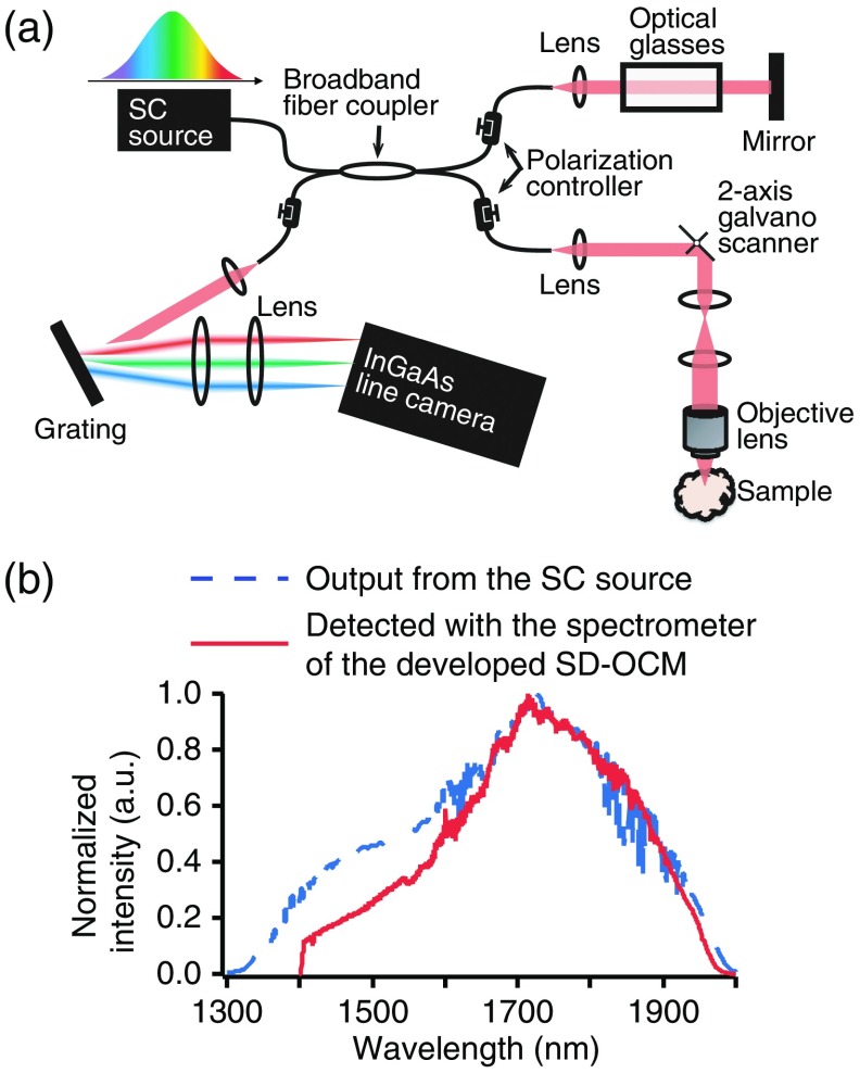
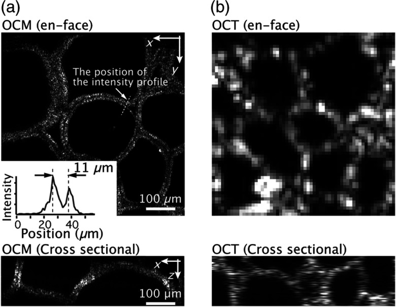
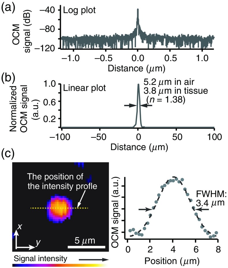
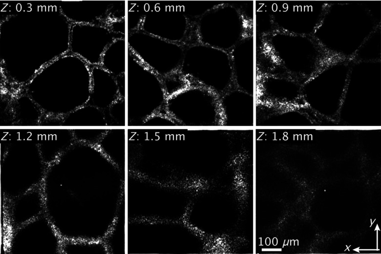
We present three-dimensional (3-D) high-resolution spectral-domain optical coherence microscopy (SD-OCM) by using a supercontinuum (SC) fiber laser source with 300-nm spectral bandwidth (full-width at half-maximum) in the 1700-nm spectral band. By using low-coherence interferometry with SC light and a confocal detection scheme, we realized lateral and axial resolutions of 3.4 and 3.8  μm in tissue (n  =  1.38), respectively. This is, to the best of our knowledge, the highest 3-D spatial resolution reported among those of Fourier-domain optical coherence imaging techniques in the 1700-nm spectral band. In our SD-OCM, to enhance the imaging depth, a full-range method was implemented, which suppressed the formation of a coherent ghost image and allowed us to set the zero-delay position inside the samples. We demonstrated the 3-D high-resolution imaging capability of 1700-nm SD-OCM through the measurement of an interference signal from a mirror surface and imaging of a single 200-nm polystyrene bead and a pig thyroid gland. Deep tissue imaging at a depth of up to 1.8 mm was also demonstrated. This is the first demonstration of 3-D high-resolution SD-OCM in the 1700-nm spectral band.

摘要

我们提出了一种基于超连续(SC)光纤激光源的三维(3-D)高分辨率光谱域光相干显微镜(SD-OCM),该激光源在 1700nm 光谱带内具有 300nm 光谱带宽(半峰全宽)。通过使用 SC 光的低相干干涉测量和共焦检测方案,我们在组织中实现了 3.4μm 的横向分辨率和 3.8μm 的轴向分辨率(n=1.38)。据我们所知,这是在 1700nm 光谱带中,傅里叶域光相干成像技术中报道的最高 3-D 空间分辨率。在我们的 SD-OCM 中,为了增强成像深度,我们采用了全范围方法,该方法抑制了相干鬼像的形成,并允许我们在样品内部设置零延迟位置。我们通过测量镜面的干涉信号以及对单个 200nm 聚苯乙烯珠和猪甲状腺的成像,展示了 1700nm SD-OCM 的 3-D 高分辨率成像能力。还展示了深度达 1.8mm 的深层组织成像。这是在 1700nm 光谱带中首次演示 3-D 高分辨率 SD-OCM。

https://cdn.ncbi.nlm.nih.gov/pmc/blobs/e1e7/6995893/30e432342431/JBO-024-070502-g004.jpg
https://cdn.ncbi.nlm.nih.gov/pmc/blobs/e1e7/6995893/7e7e57b05198/JBO-024-070502-g001.jpg
https://cdn.ncbi.nlm.nih.gov/pmc/blobs/e1e7/6995893/bcf20300673d/JBO-024-070502-g002.jpg
https://cdn.ncbi.nlm.nih.gov/pmc/blobs/e1e7/6995893/b2422f1aaece/JBO-024-070502-g003.jpg
https://cdn.ncbi.nlm.nih.gov/pmc/blobs/e1e7/6995893/30e432342431/JBO-024-070502-g004.jpg

相似文献

1
High-spatial-resolution deep tissue imaging with spectral-domain optical coherence microscopy in the 1700-nm spectral band.在 1700nm 光谱波段的光谱域光学相干显微镜中进行高空间分辨率的深层组织成像。
J Biomed Opt. 2019 Jul;24(7):1-4. doi: 10.1117/1.JBO.24.7.070502.
2
Optical coherence microscopy in 1700 nm spectral band for high-resolution label-free deep-tissue imaging.1700nm 光谱带的光学相干显微镜用于高分辨率免标记深层组织成像。
Sci Rep. 2016 Aug 22;6:31715. doi: 10.1038/srep31715.
3
Simultaneous dual-band optical coherence tomography in the spectral domain for high resolution in vivo imaging.用于高分辨率体内成像的光谱域同步双波段光学相干断层扫描技术。
Opt Express. 2009 Oct 26;17(22):19486-500. doi: 10.1364/OE.17.019486.
4
Signal-to-background ratio and lateral resolution in deep tissue imaging by optical coherence microscopy in the 1700 nm spectral band.在 1700nm 光谱波段的光学相干显微镜深层组织成像中的信号对背景比和横向分辨率。
Sci Rep. 2019 Nov 5;9(1):16041. doi: 10.1038/s41598-019-52175-9.
5
Enhancement of optical coherence microscopy in turbid media by an optical parametric amplifier.利用光学参量放大器增强浑浊介质中的光学相干显微镜成像
J Biophotonics. 2015 Jun;8(6):512-21. doi: 10.1002/jbio.201400073. Epub 2014 Sep 8.
6
Myocardial imaging using ultrahigh-resolution spectral domain optical coherence tomography.使用超高分辨率光谱域光学相干断层扫描技术进行心肌成像。
J Biomed Opt. 2016 Jun;21(6):61006. doi: 10.1117/1.JBO.21.6.061006.
7
Fiber-based photoacoustic remote sensing microscopy and spectral-domain optical coherence tomography with a dual-function 1050-nm interrogation source.基于光纤的光声远程传感显微镜和光谱域光学相干断层扫描,采用双功能 1050nm 询问光源。
J Biomed Opt. 2021 Jun;26(6). doi: 10.1117/1.JBO.26.6.066502.
8
Wide tuning range wavelength-swept laser with a single SOA at 1020 nm for ultrahigh resolution Fourier-domain optical coherence tomography.用于超高分辨率傅里叶域光学相干断层扫描的、在1020纳米处具有单个半导体光放大器的宽调谐范围波长扫描激光器。
Opt Express. 2011 Oct 24;19(22):21227-37. doi: 10.1364/OE.19.021227.
9
Extended focus depth for Fourier domain optical coherence microscopy.傅里叶域光学相干显微镜的扩展焦深
Opt Lett. 2006 Aug 15;31(16):2450-2. doi: 10.1364/ol.31.002450.
10
Compact Er:Yb:glass-laser-based supercontinuum source for high-resolution optical coherence tomography.用于高分辨率光学相干断层扫描的紧凑型掺铒镱玻璃激光超连续谱源。
Opt Express. 2008 Jul 7;16(14):10572-9. doi: 10.1364/oe.16.010572.

引用本文的文献

1
Biomimicking Atherosclerotic Vessels: A Relevant and (Yet) Sub-Explored Topic.仿生动脉粥样硬化血管:一个相关但尚未充分探索的主题。
Biomimetics (Basel). 2024 Feb 23;9(3):135. doi: 10.3390/biomimetics9030135.
2
Ultrahigh resolution spectral-domain optical coherence tomography using the 1000-1600 nm spectral band.使用1000 - 1600纳米光谱波段的超高分辨率光谱域光学相干断层扫描技术。
Biomed Opt Express. 2022 Mar 8;13(4):1939-1947. doi: 10.1364/BOE.443654. eCollection 2022 Apr 1.
3
Imaging of the skin microvascularization using spatially depolarized dynamic speckle.

本文引用的文献

1
Deep brain optical coherence tomography angiography in mice: in vivo, noninvasive imaging of hippocampal formation.在体小鼠深部脑光学相干断层血管造影:海马结构的非侵入性成像。
Sci Rep. 2018 Aug 2;8(1):11614. doi: 10.1038/s41598-018-29975-6.
2
Dual modality reflection mode optical coherence and photoacoustic microscopy using an akinetic sensor.使用无运动传感器的双模态反射模式光学相干和光声显微镜。
Opt Lett. 2017 Nov 1;42(21):4319-4322. doi: 10.1364/OL.42.004319.
3
Axial resolution and signal-to-noise ratio in deep-tissue imaging with 1.7-μm high-resolution optical coherence tomography with an ultrabroadband laser source.
利用空间去极化动态散斑成像技术对皮肤微血管进行成像。
J Biomed Opt. 2022 Apr;27(4). doi: 10.1117/1.JBO.27.4.046003.
4
Signal-to-background ratio and lateral resolution in deep tissue imaging by optical coherence microscopy in the 1700 nm spectral band.在 1700nm 光谱波段的光学相干显微镜深层组织成像中的信号对背景比和横向分辨率。
Sci Rep. 2019 Nov 5;9(1):16041. doi: 10.1038/s41598-019-52175-9.
1.7μm 超高分辨率光学相干断层成像的轴向分辨率和信噪比与超宽带激光源。
J Biomed Opt. 2017 Aug 1;22(8):85002. doi: 10.1117/1.JBO.22.8.085002.
4
Optical coherence microscopy in 1700 nm spectral band for high-resolution label-free deep-tissue imaging.1700nm 光谱带的光学相干显微镜用于高分辨率免标记深层组织成像。
Sci Rep. 2016 Aug 22;6:31715. doi: 10.1038/srep31715.
5
Noninvasive, in vivo imaging of subcortical mouse brain regions with 1.7  μm optical coherence tomography.使用1.7μm光学相干断层扫描对小鼠皮质下脑区进行无创活体成像。
Opt Lett. 2015 Nov 1;40(21):4911-4. doi: 10.1364/OL.40.004911.
6
Swept-source optical coherence tomography powered by a 1.3-μm vertical cavity surface emitting laser enables 2.3-mm-deep brain imaging in mice in vivo.由1.3微米垂直腔面发射激光器驱动的扫频源光学相干断层扫描技术能够在活体小鼠中实现2.3毫米深度的脑部成像。
J Biomed Opt. 2015 Oct;20(10):106004. doi: 10.1117/1.JBO.20.10.106004.
7
1.7-μm spectroscopic spectral-domain optical coherence tomography for imaging lipid distribution within blood vessel.用于成像血管内脂质分布的1.7微米光谱域光学相干断层扫描技术
Opt Express. 2015 Mar 9;23(5):6645-55. doi: 10.1364/OE.23.006645.
8
Development of a high power supercontinuum source in the 1.7 μm wavelength region for highly penetrative ultrahigh-resolution optical coherence tomography.用于高穿透性超高分辨率光学相干断层扫描的1.7μm波长区域高功率超连续谱源的研制。
Biomed Opt Express. 2014 Feb 26;5(3):932-43. doi: 10.1364/BOE.5.000932. eCollection 2014 Mar 1.
9
Ultrahigh speed spectral-domain optical coherence microscopy.超高速光谱域光学相干显微镜
Biomed Opt Express. 2013 Jul 1;4(8):1236-54. doi: 10.1364/BOE.4.001236. eCollection 2013.
10
Swept source optical coherence microscopy using a 1310 nm VCSEL light source.使用1310纳米垂直腔面发射激光器(VCSEL)光源的扫频源光学相干显微镜。
Opt Express. 2013 Jul 29;21(15):18021-33. doi: 10.1364/OE.21.018021.