Department of Clinical and Experimental Epilepsy, UCL Queen Square Institute of Neurology, Queen Square, London, UK.
MRI Unit, Epilepsy Society, Chalfont St Peter, Buckinghamshire, UK.
Brain. 2019 Sep 1;142(9):2670-2687. doi: 10.1093/brain/awz215.
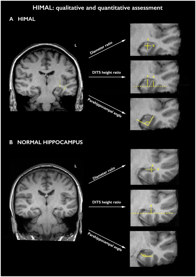
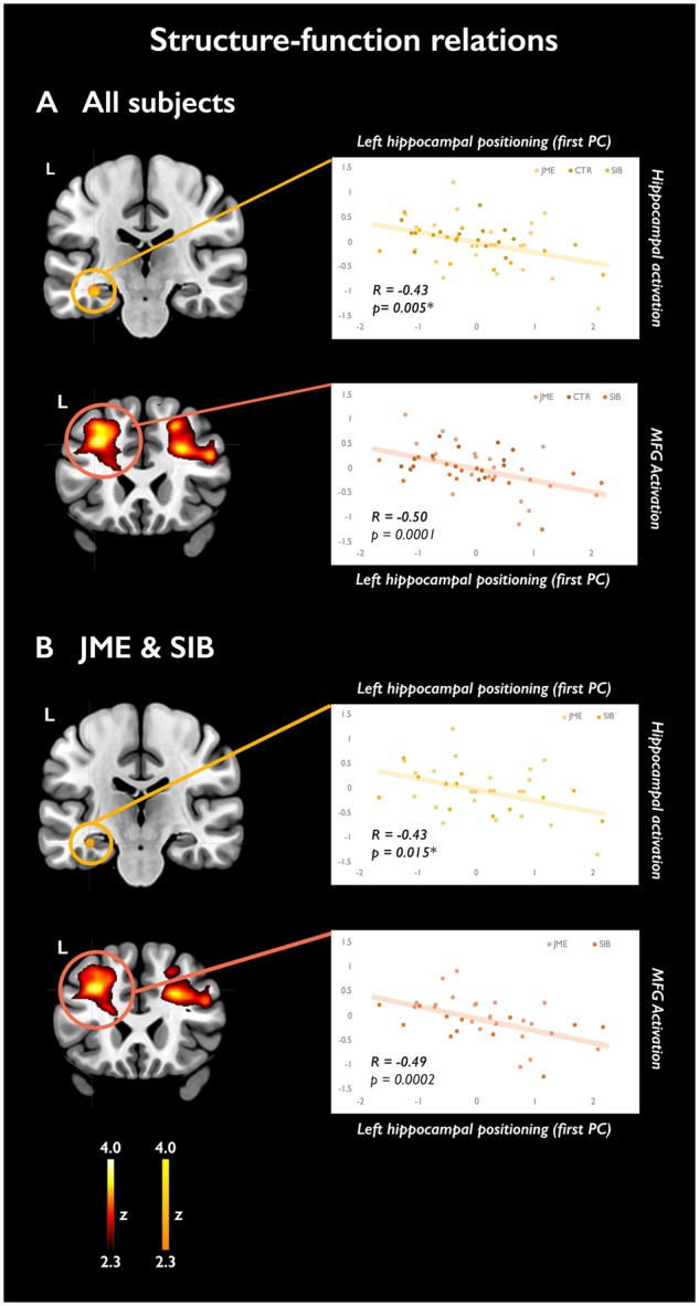
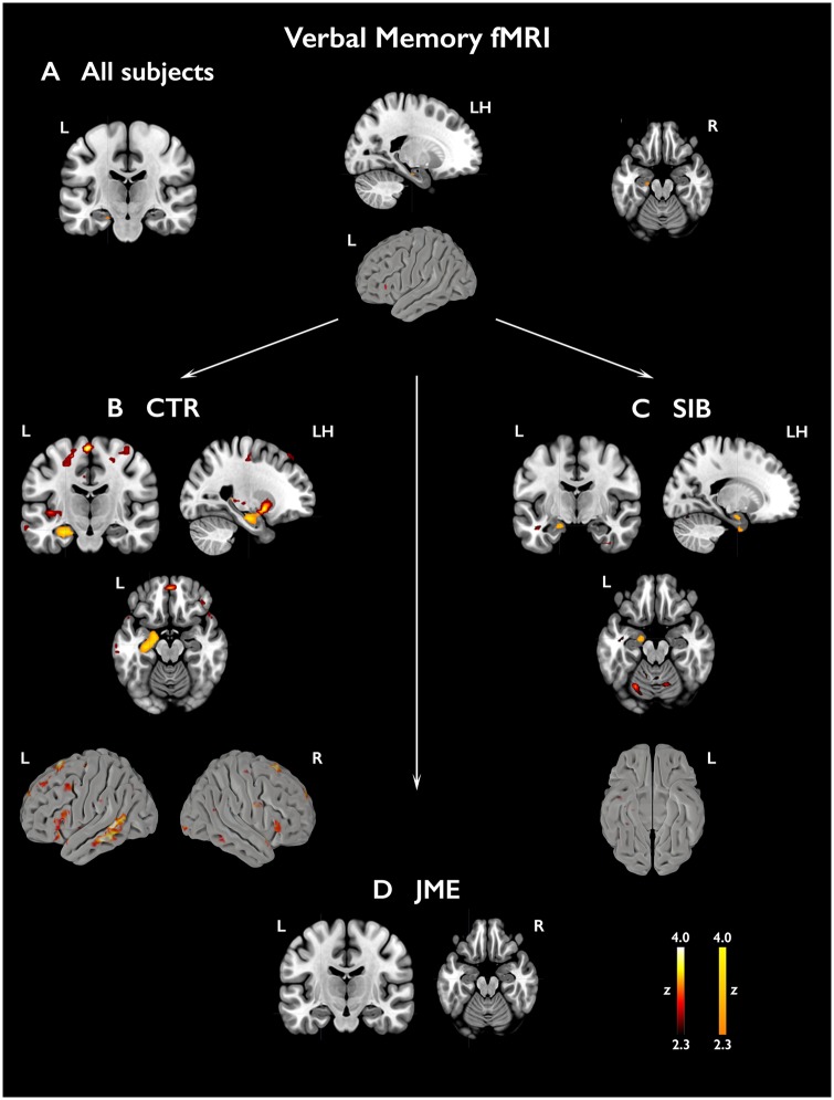
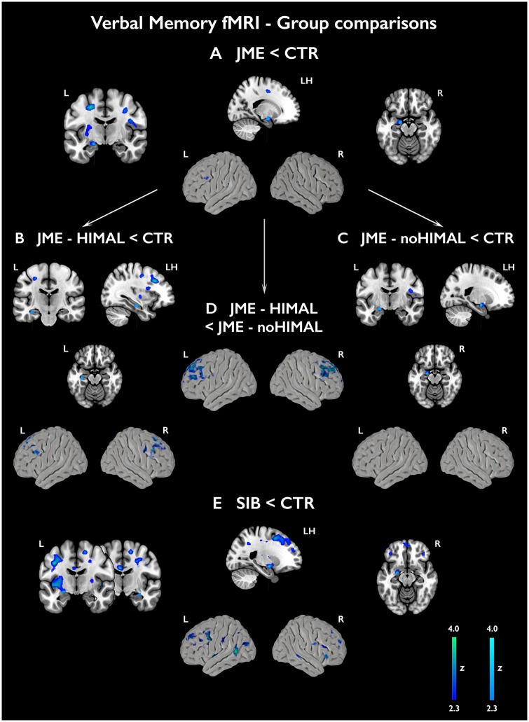
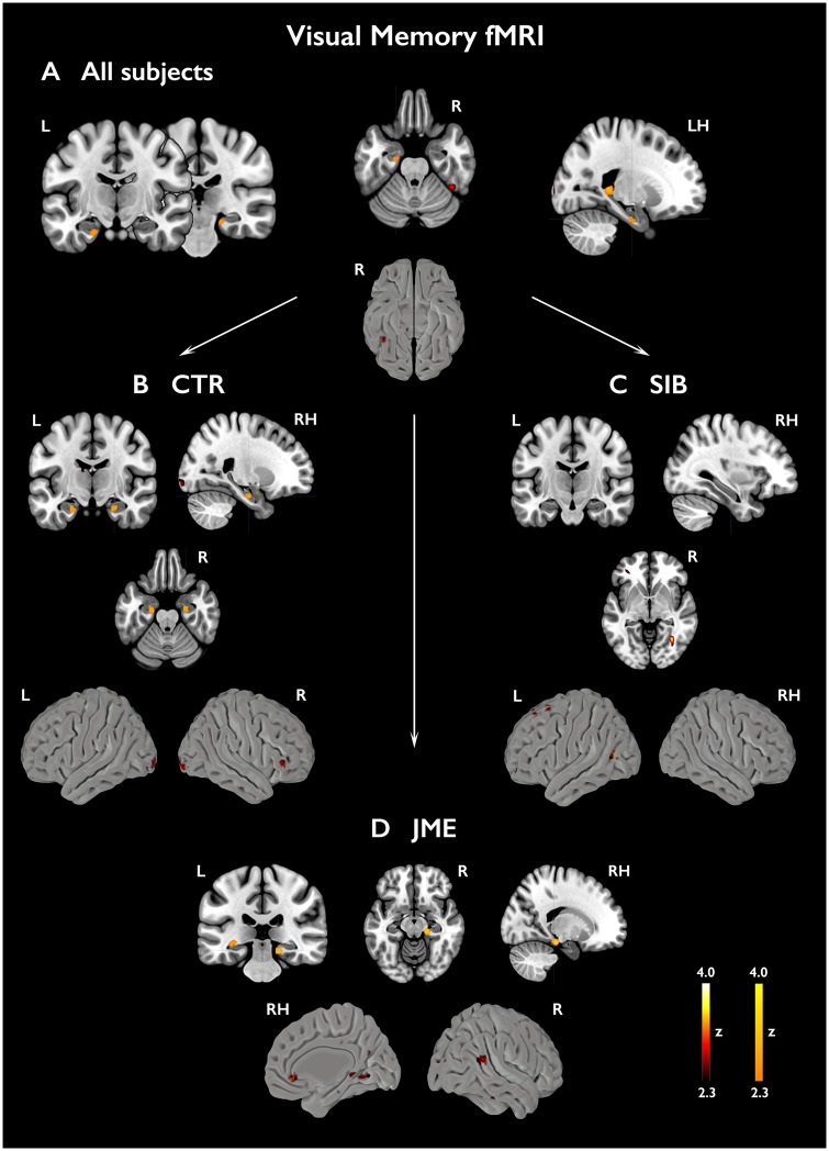
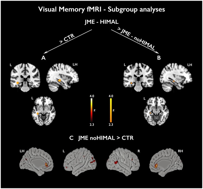
Juvenile myoclonic epilepsy is the most common genetic generalized epilepsy syndrome, characterized by a complex polygenetic aetiology. Structural and functional MRI studies demonstrated mesial or lateral frontal cortical derangements and impaired fronto-cortico-subcortical connectivity in patients and their unaffected siblings. The presence of hippocampal abnormalities and associated memory deficits is controversial, and functional MRI studies in juvenile myoclonic epilepsy have not tested hippocampal activation. In this observational study, we implemented multi-modal MRI and neuropsychological data to investigate hippocampal structure and function in 37 patients with juvenile myoclonic epilepsy, 16 unaffected siblings and 20 healthy controls, comparable for age, gender, handedness and hemispheric dominance as assessed with language laterality indices. Automated hippocampal volumetry was complemented by validated qualitative and quantitative morphological criteria to detect hippocampal malrotation, assumed to represent a neurodevelopmental marker. Neuropsychological measures of verbal and visuo-spatial learning and an event-related verbal and visual memory functional MRI paradigm addressed mesiotemporal function. We detected a reduction of mean left hippocampal volume in patients and their siblings compared with controls (P < 0.01). Unilateral or bilateral hippocampal malrotation was identified in 51% of patients and 50% of siblings, against 15% of controls (P < 0.05). For bilateral hippocampi, quantitative markers of verticalization had significantly larger values in patients and siblings compared with controls (P < 0.05). In the patient subgroup, there was no relationship between structural measures and age at disease onset or degree of seizure control. No overt impairment of verbal and visual memory was identified with neuropsychological tests. Functional mapping highlighted atypical patterns of hippocampal activation, pointing to abnormal recruitment during verbal encoding in patients and their siblings [P < 0.05, familywise error (FWE)-corrected]. Subgroup analyses indicated distinct profiles of hypoactivation along the hippocampal long axis in juvenile myoclonic epilepsy patients with and without malrotation; patients with malrotation also exhibited reduced frontal recruitment for verbal memory, and more pronounced left posterior hippocampal involvement for visual memory. Linear models across the entire study cohort indicated significant associations between morphological markers of hippocampal positioning and hippocampal activation for verbal items (all P < 0.05, FWE-corrected). We demonstrate abnormalities of hippocampal volume, shape and positioning in patients with juvenile myoclonic epilepsy and their siblings, which are associated with reorganization of function and imply an underlying neurodevelopmental mechanism with expression during the prenatal stage. Co-segregation of abnormal hippocampal morphology in patients and their siblings is suggestive of a genetic imaging phenotype, independent of disease activity, and can be construed as a novel endophenotype of juvenile myoclonic epilepsy.
青少年肌阵挛性癫痫是最常见的遗传性全面性癫痫综合征,其病因复杂,具有遗传异质性。结构和功能磁共振成像研究表明,患者及其无癫痫发作的兄弟姐妹存在额叶皮质内侧或外侧的紊乱和额皮质下连接的损伤。海马异常和相关记忆缺陷的存在存在争议,并且青少年肌阵挛性癫痫的功能磁共振成像研究尚未测试海马激活。在这项观察性研究中,我们采用多模态 MRI 和神经心理学数据来研究 37 名青少年肌阵挛性癫痫患者、16 名无癫痫发作的兄弟姐妹和 20 名健康对照者的海马结构和功能,这些患者在年龄、性别、利手性和语言偏侧性指数评估的优势半球方面具有可比性。自动海马容积测量法补充了经过验证的定性和定量形态学标准,以检测假定代表神经发育标志物的海马旋转不良。内侧颞叶功能的神经心理学测量包括言语和视觉空间学习以及事件相关的言语和视觉记忆功能磁共振成像范式。我们发现患者及其兄弟姐妹的左侧海马体体积平均值较对照组减小(P < 0.01)。51%的患者和 50%的兄弟姐妹存在单侧或双侧海马旋转不良,而对照组为 15%(P < 0.05)。对于双侧海马,垂直化的定量标志物在患者和兄弟姐妹中的值明显大于对照组(P < 0.05)。在患者亚组中,结构测量与疾病发病年龄或癫痫发作控制程度之间没有关系。神经心理学测试未发现言语和视觉记忆明显受损。功能映射突出了海马激活的异常模式,表明患者及其兄弟姐妹在言语编码过程中存在异常招募[P < 0.05,家族性错误(FWE)校正]。亚组分析表明,在有或没有旋转不良的青少年肌阵挛性癫痫患者中,沿海马长轴存在不同的低激活谱;旋转不良的患者在言语记忆中额叶招募减少,视觉记忆中左后海马参与程度增加。整个研究队列的线性模型表明,海马定位的形态学标志物与言语项目的海马激活之间存在显著关联(所有 P < 0.05,FWE 校正)。我们在青少年肌阵挛性癫痫患者及其兄弟姐妹中发现了海马体积、形状和定位的异常,这些异常与功能重组有关,并暗示了一种潜在的神经发育机制,该机制在产前阶段表达。患者及其兄弟姐妹中异常海马形态的共分离提示存在遗传影像学表型,与疾病活动无关,可作为青少年肌阵挛性癫痫的新内表型。