Visceral Surgery Department, Dupuytren University Hospital, Limoges, France.
University Limoges, CNRS, XLIM, UMR 7252, Limoges, France.
PLoS One. 2019 Jul 31;14(7):e0220360. doi: 10.1371/journal.pone.0220360. eCollection 2019.
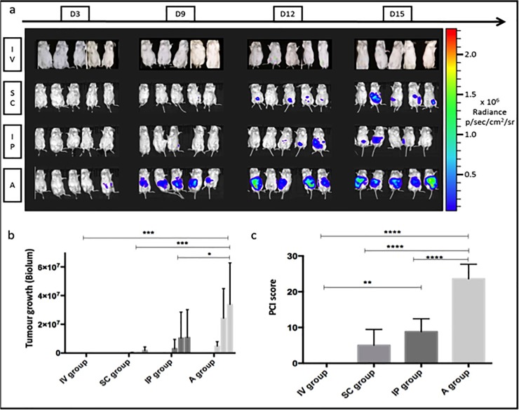
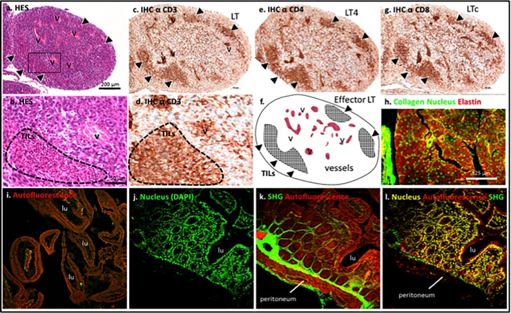
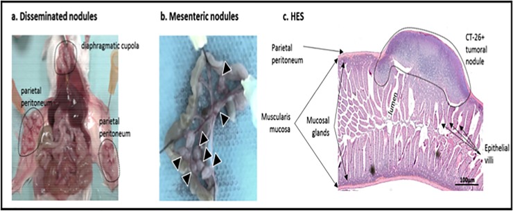
The development of cancer mouse models is still needed for the identification and preclinical validation of novel therapeutic targets in colorectal cancer, which is the third leading cause of cancer-related deaths in Europe. The purpose of this study was to determine the most accurate tumour cell injection method to obtain suitable peritoneal metastasis (PM) for subsequent therapeutic treatments. Here, we grafted murine colon carcinoma CT-26 cells expressing luciferase into immunocompetent BALB-c mice by intravenous injection (IV group), subcutaneous injection (SC group), intraperitoneal injection after peritoneal scratching (A group) or intraperitoneal injection alone (IP group). Tumour growth was monitored by bioluminescence during the first 15 days post-grafting. The peritoneal carcinomatosis index was evaluated macroscopically, histology, immunohistochemistry and multiphoton microscopy were performed in peritoneal tumour tissue. Upon implantation, no tumour growth was observed in the IV group, similar to the non-injected group. Both the IP and SC groups showed intermediate growth rates, but the SC group produced only a single subcutaneous nodule. The A group exhibited the highest tumour growth at 15 days post-surgery. Anatomic and histologic analyses corroborated the existence of various tumour nodules, and multiphoton microscopy was used to evaluate tumour fibrosis-infiltrating cells in a non-pathologic peritoneum. In conclusion, limited PM was obtained by IP injection, whereas IP injection after peritoneal scratching led to an extensive PM murine model for evaluating new therapeutics.
癌症小鼠模型的发展仍然是必要的,以鉴定和验证结直肠癌的新治疗靶点,结直肠癌是欧洲癌症相关死亡的第三大原因。本研究的目的是确定最准确的肿瘤细胞注射方法,以获得适合后续治疗的腹膜转移(PM)。在这里,我们将表达荧光素酶的鼠结肠癌细胞 CT-26 经静脉注射(IV 组)、皮下注射(SC 组)、腹膜划痕后腹腔内注射(A 组)或单纯腹腔内注射(IP 组)移植到免疫功能正常的 BALB/c 小鼠体内。在移植后 15 天内通过生物发光监测肿瘤生长。用肉眼、组织学、免疫组织化学和多光子显微镜观察腹膜肿瘤组织评估腹膜癌转移指数。在植入时,IV 组没有观察到肿瘤生长,与未注射组相似。IP 组和 SC 组的生长速度均为中等,但 SC 组仅产生单个皮下结节。A 组在手术后 15 天表现出最高的肿瘤生长。解剖学和组织学分析证实存在各种肿瘤结节,多光子显微镜用于评估非病理性腹膜中肿瘤纤维化浸润细胞。总之,IP 注射获得了有限的 PM,而腹膜划痕后的 IP 注射导致了广泛的 PM 小鼠模型,用于评估新的治疗方法。