Department of Neurology, Johns Hopkins University School of Medicine, Baltimore, MD, 21205, USA.
Department of Neuroscience, Johns Hopkins University, Baltimore, MD, USA.
Acta Neuropathol Commun. 2019 Jul 31;7(1):125. doi: 10.1186/s40478-019-0767-6.
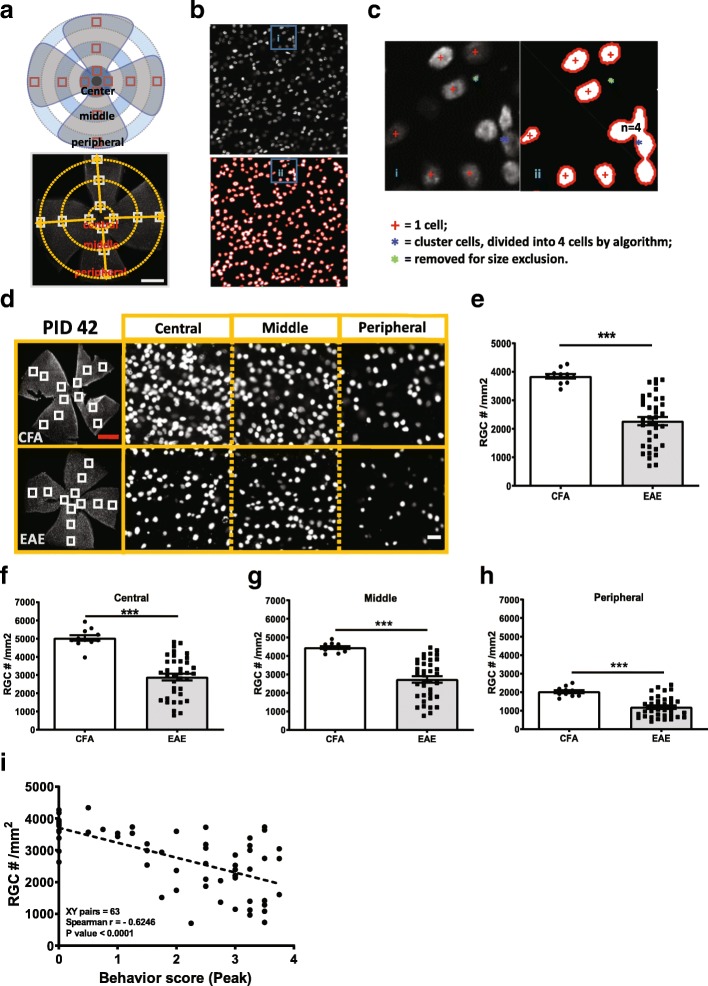
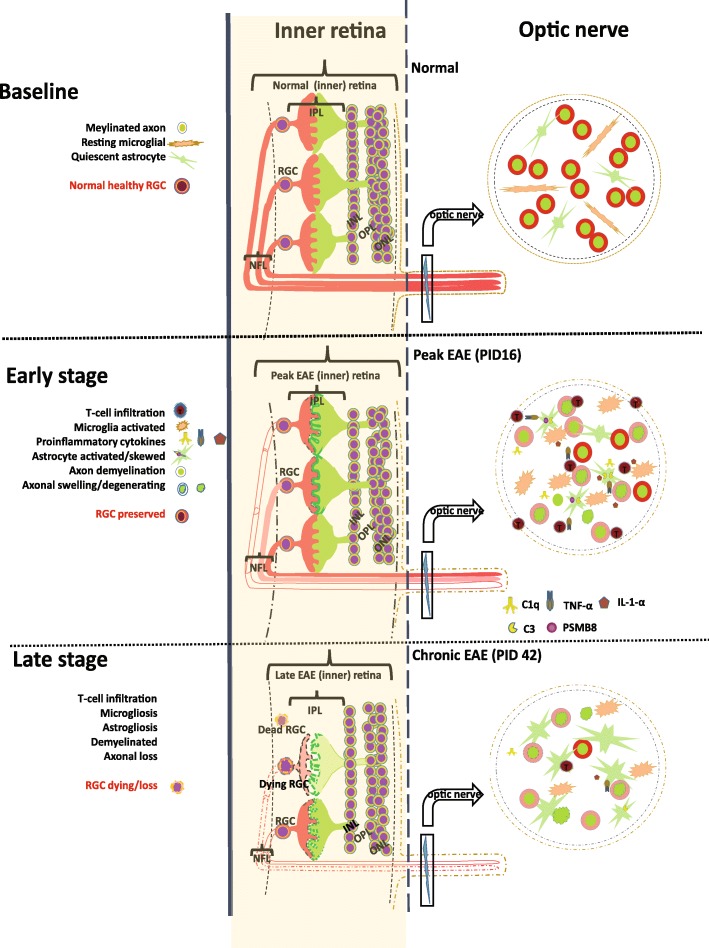
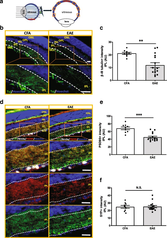
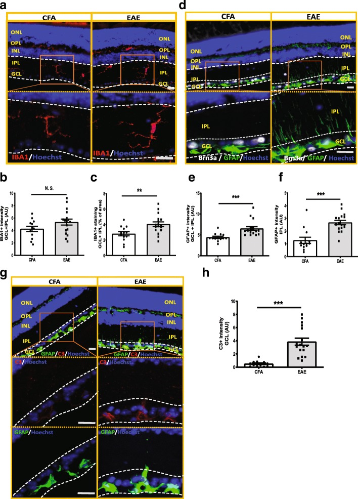
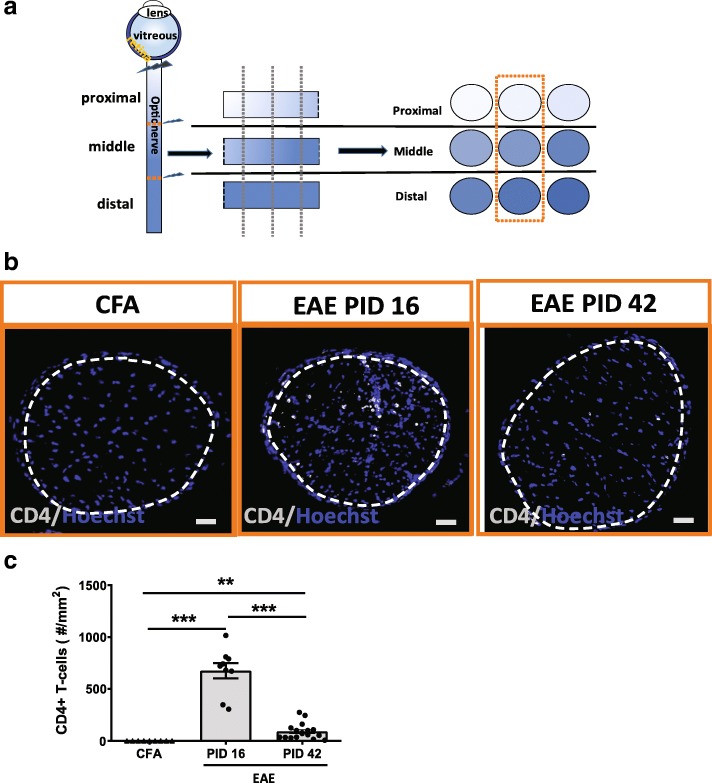
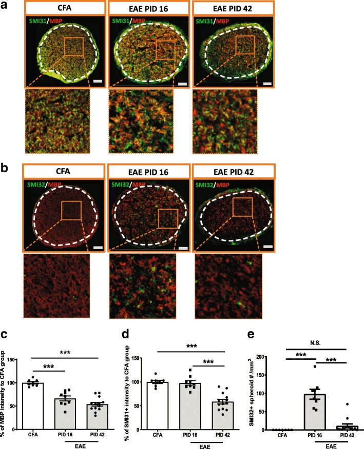
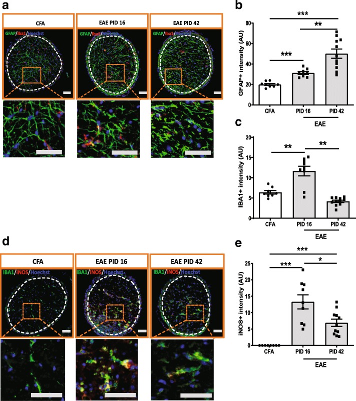
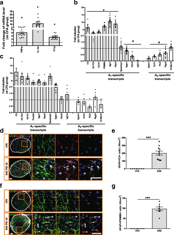
The animal model experimental autoimmune encephalomyelitis (EAE) has been used extensively in the past to test mechanisms that target peripheral immune cells for treatment of multiple sclerosis (MS). While there have been some notable successes in relapsing MS, the development of therapies for progressive multiple sclerosis (MS) has been hampered by lack of an appropriate animal model. Further, the mechanisms underlying CNS inflammation and neuronal injury remain incompletely elucidated. It is known that the MOG EAE mouse model does not have insidious behavioral progression as occurs in people with MS, but there is significant neuronal and axonal injury in EAE, as a result of the inflammation. In the present study, we describe the time course of glial activation and retinal neurodegeneration in the EAE model, and highlight the utility of studying the anterior visual pathway for modeling mechanisms of neuronal injury that may recapitulate critical aspects of the pathology described in people with MS following optic neuritis and subclinical optic neuropathy. We show that A1 neurotoxic astrocytes are prevalent in optic nerve tissue and retina, and are associated with subsequent RGC loss in the most commonly used form of the EAE model induced by MOG peptide in C57/B6 mice. We developed a semi-automatic method to quantify retinal ganglion cells (RGC) and show that RGCs remain intact at peak EAE (PID 16) but are significantly reduced in late EAE (PID 42). Postsynaptic proteins and neurites were also compromised in the retina of late EAE mice. The retinal pathology manifests weeks after the microglial and astrocyte activation, which were prominent in optic nerve tissues at PID 16. Microglia expressed iNOS and had increased gene expression of C1q, TNF-α, and IL-1α. Astrocytes expressed high levels of complement component 3 and other genes associated with A1 neurotoxic astrocytes. Our data suggest that EAE can be used to study the pathobiology of optic neuropathy and to examine the preclinical neuroprotective effects of drugs that target activation of neurotoxic A1 astrocytes.
实验性自身免疫性脑脊髓炎(EAE)动物模型在过去被广泛用于测试针对外周免疫细胞的治疗多发性硬化症(MS)的机制。虽然在复发性 MS 方面取得了一些显著的成功,但由于缺乏合适的动物模型,进展性多发性硬化症(MS)的治疗方法的发展受到了阻碍。此外,中枢神经系统炎症和神经元损伤的机制仍未完全阐明。已知 MOG EAE 小鼠模型没有 MS 患者中发生的隐匿性行为进展,但 EAE 中有明显的神经元和轴突损伤,这是炎症的结果。在本研究中,我们描述了 EAE 模型中神经胶质激活和视网膜神经退行性变的时间过程,并强调了研究前视通路对于模拟可能再现 MS 患者视神经炎和亚临床视神经病变后病理学关键方面的神经元损伤机制的效用。我们表明,A1 神经毒性星形胶质细胞在视神经组织和视网膜中普遍存在,并且与最常用的 C57/B6 小鼠 MOG 肽诱导的 EAE 模型中随后的 RGC 损失相关。我们开发了一种半自动方法来量化视网膜神经节细胞(RGC),并表明 RGC 在 EAE 高峰期(PID 16)保持完整,但在晚期 EAE(PID 42)中显著减少。晚期 EAE 小鼠的视网膜中也存在突触后蛋白和神经突受损。视网膜病理学在小胶质细胞和星形胶质细胞激活后数周表现出来,在 PID 16 时在视神经组织中非常明显。小胶质细胞表达 iNOS,并增加了 C1q、TNF-α 和 IL-1α 的基因表达。星形胶质细胞表达高水平的补体成分 3 和其他与 A1 神经毒性星形胶质细胞相关的基因。我们的数据表明,EAE 可用于研究视神经病变的病理生物学,并检查针对激活神经毒性 A1 星形胶质细胞的药物的临床前神经保护作用。