Baldinger-Melich Pia, Urquijo Castro Maria F, Seiger René, Ruef Anne, Dwyer Dominic B, Kranz Georg S, Klöbl Manfred, Kambeitz Joseph, Kaufmann Ulrike, Windischberger Christian, Kasper Siegfried, Falkai Peter, Lanzenberger Rupert, Koutsouleris Nikolaos
Department of Psychiatry and Psychotherapy, Clinical Division of General Psychiatry, Medical University of Vienna, Vienna, Austria.
Neuroimaging Labs (NIL) PET, MRI, EEG, TMS and Chemical Lab, Department of Psychiatry and Psychotherapy, Clinical Division of General Psychiatry, Medical University of Vienna, Vienna, Austria.
Cereb Cortex. 2020 Mar 14;30(3):1345-1356. doi: 10.1093/cercor/bhz170.
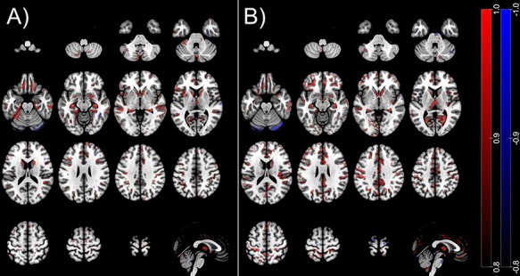
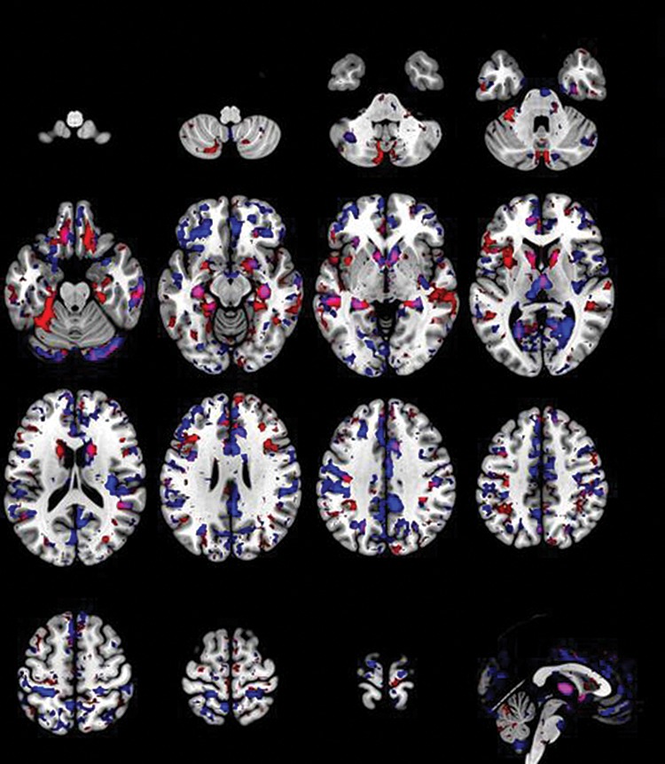
Univariate analyses of structural neuroimaging data have produced heterogeneous results regarding anatomical sex- and gender-related differences. The current study aimed at delineating and cross-validating brain volumetric surrogates of sex and gender by comparing the structural magnetic resonance imaging data of cis- and transgender subjects using multivariate pattern analysis. Gray matter (GM) tissue maps of 29 transgender men, 23 transgender women, 35 cisgender women, and 34 cisgender men were created using voxel-based morphometry and analyzed using support vector classification. Generalizability of the models was estimated using repeated nested cross-validation. For external validation, significant models were applied to hormone-treated transgender subjects (n = 32) and individuals diagnosed with depression (n = 27). Sex was identified with a balanced accuracy (BAC) of 82.6% (false discovery rate [pFDR] < 0.001) in cisgender, but only with 67.5% (pFDR = 0.04) in transgender participants indicating differences in the neuroanatomical patterns associated with sex in transgender despite the major effect of sex on GM volume irrespective of the self-identification as a woman or man. Gender identity and gender incongruence could not be reliably identified (all pFDR > 0.05). The neuroanatomical signature of sex in cisgender did not interact with depressive features (BAC = 74.7%) but was affected by hormone therapy when applied in transgender women (P < 0.001).
对结构神经影像数据的单变量分析在解剖学上与性别相关的差异方面产生了异质性结果。本研究旨在通过使用多变量模式分析比较顺性别和跨性别受试者的结构磁共振成像数据,来描绘和交叉验证性别和性别的脑容量替代指标。使用基于体素的形态计量学创建了29名跨性别男性、23名跨性别女性、35名顺性别女性和34名顺性别男性的灰质(GM)组织图,并使用支持向量分类进行分析。使用重复嵌套交叉验证估计模型的可推广性。为了进行外部验证,将显著模型应用于接受激素治疗的跨性别受试者(n = 32)和被诊断患有抑郁症的个体(n = 27)。在顺性别者中,性别识别的平衡准确率(BAC)为82.6%(错误发现率[pFDR]<0.001),但在跨性别参与者中仅为67.5%(pFDR = 0.04),这表明尽管性别对GM体积有主要影响,无论自我认同为女性还是男性,跨性别者中与性别相关的神经解剖模式存在差异。性别认同和性别不一致无法可靠识别(所有pFDR>0.05)。顺性别者中性别相关的神经解剖特征与抑郁特征没有相互作用(BAC = 74.7%),但在跨性别女性中应用时会受到激素治疗的影响(P<0.001)。