• 文献检索
  • 文档翻译
  • 深度研究
  • 学术资讯
  • Suppr Zotero 插件Zotero 插件
  • 邀请有礼
  • 套餐&价格
  • 历史记录
应用&插件
Suppr Zotero 插件Zotero 插件浏览器插件Mac 客户端Windows 客户端微信小程序
定价
高级版会员购买积分包购买API积分包
服务
文献检索文档翻译深度研究API 文档MCP 服务
关于我们
关于 Suppr公司介绍联系我们用户协议隐私条款
关注我们

Suppr 超能文献

核心技术专利:CN118964589B侵权必究
粤ICP备2023148730 号-1Suppr @ 2026

文献检索

告别复杂PubMed语法,用中文像聊天一样搜索,搜遍4000万医学文献。AI智能推荐,让科研检索更轻松。

立即免费搜索

文件翻译

保留排版,准确专业,支持PDF/Word/PPT等文件格式,支持 12+语言互译。

免费翻译文档

深度研究

AI帮你快速写综述,25分钟生成高质量综述,智能提取关键信息,辅助科研写作。

立即免费体验

UVA 辐射暴露后视网膜色素上皮细胞的蛋白质组学分析。

Proteomic analysis of retinal pigment epithelium cells after exposure to UVA radiation.

机构信息

Department of Ophthalmology, Kaohsiung Veterans General Hospital, Kaohsiung, Taiwan.

School of Medicine, National Yang-Ming University, Taipei, Taiwan.

出版信息

BMC Ophthalmol. 2019 Aug 2;19(1):168. doi: 10.1186/s12886-019-1151-9.

DOI:10.1186/s12886-019-1151-9
PMID:31375076
原文链接:https://pmc.ncbi.nlm.nih.gov/articles/PMC6679551/
Abstract

BACKGROUND

Age-related macular degeneration (AMD) is the primary cause of blindness and severe vision loss in developed countries and is responsible for 8.7% of blindness globally. Ultraviolet radiation can induce DNA breakdown, produce reactive oxygen species, and has been implicated as a risk factor for AMD. This study investigated the effects of UVA radiation on Human retinal pigment epithelial cell (ARPE-19) growth and protein expression.

METHODS

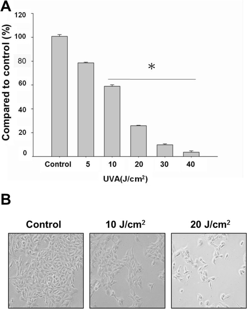
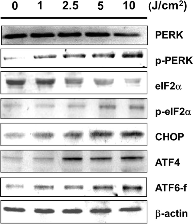
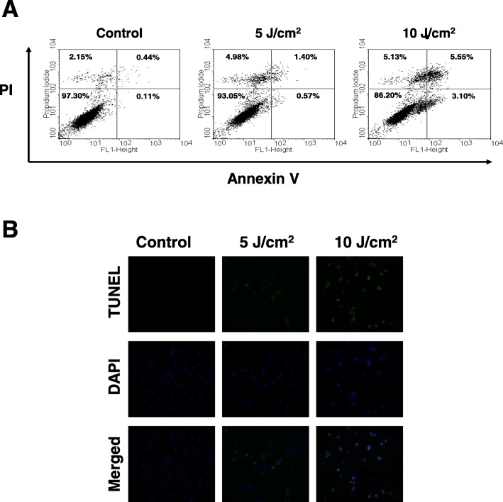
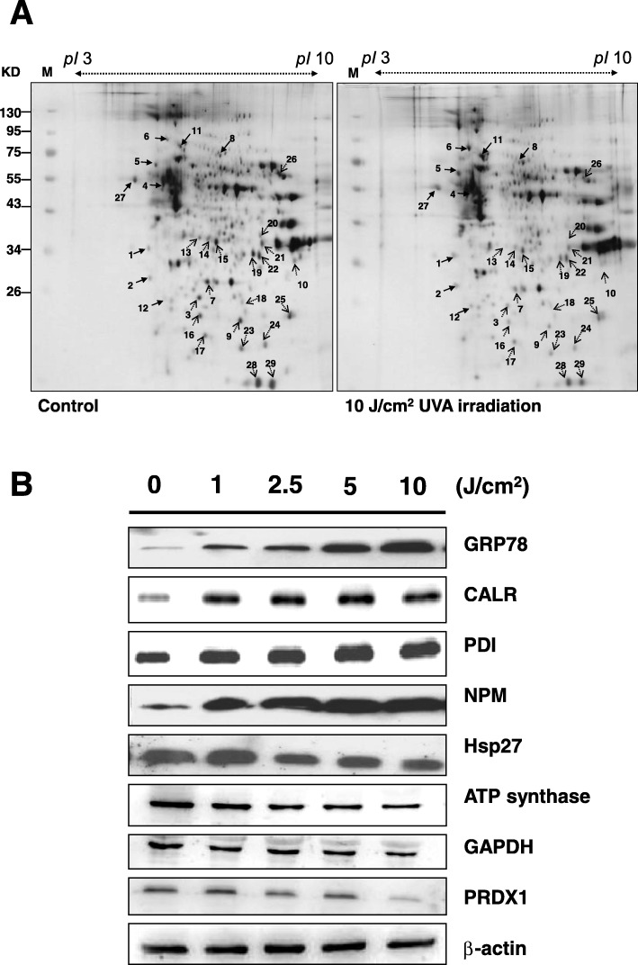
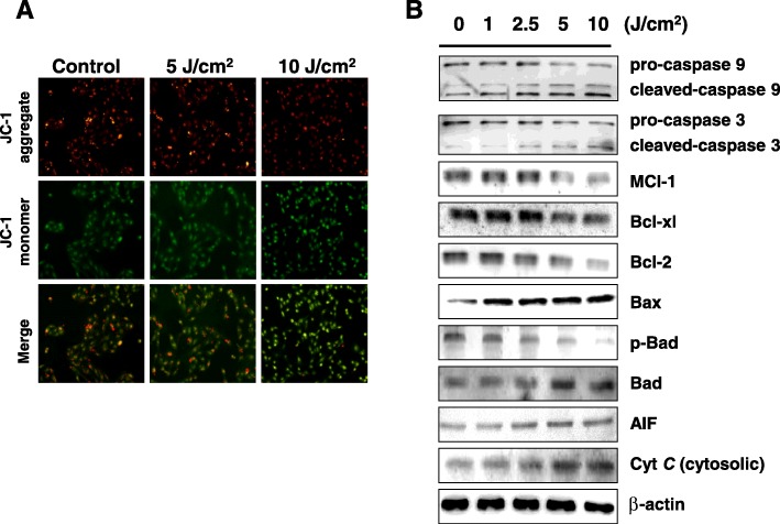
ARPE-19 cells were irradiated with a UVA lamp at different doses (5, 10, 20, 30 and 40 J/cm) from 10 cm. Cell viability was determined by MTT assay. Visual inspection was first achieved with inverted light microscopy and then the DeadEnd™ Fluorometric TUNEL System was used to observe nuclear DNA fragmentation. Flow cytometry based-Annexin V-FITC/PI double-staining was used to further quantify cellular viability. Mitochondrial membrane potential was assessed with JC-1 staining. 2D electrophoresis maps of exposed cells were compared to nonexposed cells and gel images analyzed with PDQuest 2-D Analysis Software. Spots with greater than a 1.5-fold difference were selected for LC-MS/MS analysis and some confirmed by western blot. We further investigated whether caspase activation, apoptotic-related mitochondrial proteins, and regulators of ER stress sensors were involved in UVA-induced apoptosis.

RESULTS

We detected 29 differentially expressed proteins (9 up-regulated and 20 down-regulated) in the exposed cells. Some of these proteins such as CALR, GRP78, NPM, Hsp27, PDI, ATP synthase subunit alpha, PRDX1, and GAPDH are associated with anti-proliferation, induction of apoptosis, and oxidative-stress protection. We also detected altered protein expression levels among caspases (caspase 3 and 9) and in the mitochondrial (cytosolic cytochrome C, AIF, Mcl-1, Bcl-2, Bcl-xl, Bax, Bad, and p-Bad) and ER stress-related (p-PERK, p-eIF2α, ATF4 and CHOP) apoptotic pathways.

CONCLUSIONS

UVA irradiation suppressed the proliferation of ARPE-19 cells in a dose-dependent manner, caused quantitative loses in transmembrane potential (ΔΨm), and induced both early and late apoptosis.

摘要

背景

年龄相关性黄斑变性(AMD)是发达国家致盲和严重视力丧失的主要原因,也是全球 8.7%失明的原因。紫外线辐射可导致 DNA 断裂,产生活性氧,并被认为是 AMD 的一个风险因素。本研究探讨了 UVA 辐射对人视网膜色素上皮细胞(ARPE-19)生长和蛋白表达的影响。

方法

ARPE-19 细胞用 UVA 灯从 10cm 处照射不同剂量(5、10、20、30 和 40 J/cm)。通过 MTT 测定法测定细胞活力。首先通过倒置相差显微镜进行目视检查,然后使用 DeadEnd™荧光 TUNEL 系统观察核 DNA 片段化。使用 Annexin V-FITC/PI 双染色的流式细胞术进一步定量细胞活力。使用 JC-1 染色评估线粒体膜电位。将暴露细胞的 2D 电泳图谱与未暴露细胞进行比较,并使用 PDQuest 2-D 分析软件分析凝胶图像。选择差异大于 1.5 倍的斑点进行 LC-MS/MS 分析,并通过 Western blot 验证一些结果。我们进一步研究了 caspase 激活、凋亡相关的线粒体蛋白以及内质网应激传感器的调节剂是否参与了 UVA 诱导的细胞凋亡。

结果

我们在暴露细胞中检测到 29 种差异表达蛋白(9 种上调和 20 种下调)。其中一些蛋白,如 CALR、GRP78、NPM、Hsp27、PDI、ATP 合酶亚基α、PRDX1 和 GAPDH,与抗增殖、诱导凋亡和氧化应激保护有关。我们还检测到细胞色素 C、AIF、Mcl-1、Bcl-2、Bcl-xl、Bax、Bad 和 p-Bad 等 caspase(caspase 3 和 9)以及线粒体(细胞质)和内质网应激相关(p-PERK、p-eIF2α、ATF4 和 CHOP)凋亡途径中的蛋白表达水平改变。

结论

UVA 照射以剂量依赖性方式抑制 ARPE-19 细胞的增殖,导致跨膜电位(ΔΨm)定量损失,并诱导早期和晚期凋亡。

https://cdn.ncbi.nlm.nih.gov/pmc/blobs/dc33/6679551/a0374348a675/12886_2019_1151_Fig5_HTML.jpg
https://cdn.ncbi.nlm.nih.gov/pmc/blobs/dc33/6679551/f691e4f58aa4/12886_2019_1151_Fig1_HTML.jpg
https://cdn.ncbi.nlm.nih.gov/pmc/blobs/dc33/6679551/178a8c2843d5/12886_2019_1151_Fig2_HTML.jpg
https://cdn.ncbi.nlm.nih.gov/pmc/blobs/dc33/6679551/e089b286fd37/12886_2019_1151_Fig3_HTML.jpg
https://cdn.ncbi.nlm.nih.gov/pmc/blobs/dc33/6679551/72552d22d424/12886_2019_1151_Fig4_HTML.jpg
https://cdn.ncbi.nlm.nih.gov/pmc/blobs/dc33/6679551/a0374348a675/12886_2019_1151_Fig5_HTML.jpg

相似文献

1
Proteomic analysis of retinal pigment epithelium cells after exposure to UVA radiation.UVA 辐射暴露后视网膜色素上皮细胞的蛋白质组学分析。
BMC Ophthalmol. 2019 Aug 2;19(1):168. doi: 10.1186/s12886-019-1151-9.
2
Expression of early and late cellular damage markers by ARPE-19 cells following prolonged treatment with UV-A radiation.用UV-A辐射长时间处理后ARPE-19细胞中早期和晚期细胞损伤标志物的表达。
Mol Med Rep. 2016 Oct;14(4):3485-9. doi: 10.3892/mmr.2016.5649. Epub 2016 Aug 19.
3
Activation of the ERK1/2-MAPK Signaling Pathway by Complement Serum in UV-POS-Pretreated ARPE-19 Cells.补体血清对紫外线预处理的ARPE-19细胞中ERK1/2-MAPK信号通路的激活作用
Ophthalmologica. 2018;239(4):215-224. doi: 10.1159/000486404. Epub 2018 Feb 27.
4
Involvement of endoplasmic reticulum stress in all-trans-retinal-induced retinal pigment epithelium degeneration.内质网应激参与全反式视黄醛诱导的视网膜色素上皮细胞变性。
Toxicol Sci. 2015 Jan;143(1):196-208. doi: 10.1093/toxsci/kfu223. Epub 2014 Oct 20.
5
Stress responses of human retinal pigment epithelial cells to glyoxal.人视网膜色素上皮细胞对乙二醛的应激反应。
Graefes Arch Clin Exp Ophthalmol. 2016 Dec;254(12):2361-2372. doi: 10.1007/s00417-016-3463-2. Epub 2016 Aug 12.
6
The Role of Oxidative Stress and Autophagy in Blue-Light-Induced Damage to the Retinal Pigment Epithelium in Zebrafish In Vitro and In Vivo.氧化应激和自噬在体外和体内蓝光诱导斑马鱼视网膜色素上皮损伤中的作用。
Int J Mol Sci. 2021 Jan 29;22(3):1338. doi: 10.3390/ijms22031338.
7
Protective effect of canolol from oxidative stress-induced cell damage in ARPE-19 cells via an ERK mediated antioxidative pathway.菜子醇通过ERK介导的抗氧化途径对氧化应激诱导的ARPE-19细胞损伤的保护作用。
Mol Vis. 2011;17:2040-8. Epub 2011 Jul 27.
8
Comparative transcriptomic analysis reveals adriamycin-induced apoptosis via p53 signaling pathway in retinal pigment epithelial cells.比较转录组学分析揭示阿霉素诱导视网膜色素上皮细胞凋亡的 p53 信号通路。
J Zhejiang Univ Sci B. 2018;19(12):895-909. doi: 10.1631/jzus.B1800408.
9
Expression of endoplasmic reticulum stress markers GRP78 and CHOP induced by oxidative stress in blue light-mediated damage of A2E-containing retinal pigment epithelium cells.蓝光介导含A2E的视网膜色素上皮细胞损伤中氧化应激诱导的内质网应激标志物GRP78和CHOP的表达。
Ophthalmic Res. 2014;52(4):224-33. doi: 10.1159/000363387. Epub 2014 Nov 12.
10
PRDX1 exerts a photoprotection effect by inhibiting oxidative stress and regulating MAPK signaling on retinal pigment epithelium.PRDX1 通过抑制氧化应激和调节 MAPK 信号转导对视网膜色素上皮发挥光保护作用。
BMC Ophthalmol. 2024 Jun 6;24(1):237. doi: 10.1186/s12886-024-03489-4.

引用本文的文献

1
PRDX1 exerts a photoprotection effect by inhibiting oxidative stress and regulating MAPK signaling on retinal pigment epithelium.PRDX1 通过抑制氧化应激和调节 MAPK 信号转导对视网膜色素上皮发挥光保护作用。
BMC Ophthalmol. 2024 Jun 6;24(1):237. doi: 10.1186/s12886-024-03489-4.
2
Crosstalk Between Microglia and Müller Glia in the Age-Related Macular Degeneration: Role and Therapeutic Value of Neuroinflammation.小胶质细胞与年龄相关性黄斑变性中 Müller 胶质细胞的串扰:神经炎症的作用和治疗价值。
Aging Dis. 2024 May 7;15(3):1132-1154. doi: 10.14336/AD.2023.0823-3.
3
UVA/UVB Irradiation Exerts a Distinct Phototoxic Effect on Human Keratinocytes Compared to Human Malignant Melanoma Cells.

本文引用的文献

1
Influence of standard corneal cross-linking in keratoconus patients on macular profile.圆锥角膜患者中标准角膜交联对黄斑形态的影响。
J Curr Ophthalmol. 2018 Jun 29;30(4):330-336. doi: 10.1016/j.joco.2018.06.003. eCollection 2018 Dec.
2
Macular phototoxicity after corneal cross-linking.角膜交联术后黄斑光毒性
Clin Ophthalmol. 2018 Sep 17;12:1801-1807. doi: 10.2147/OPTH.S176025. eCollection 2018.
3
Conventional vs. pulsed-light accelerated corneal collagen cross-linking for the treatment of progressive keratoconus: 12-month results from a prospective study.
与人类恶性黑色素瘤细胞相比,UVA/UVB照射对人类角质形成细胞具有明显的光毒性作用。
Life (Basel). 2023 May 8;13(5):1144. doi: 10.3390/life13051144.
4
Serum Autoantibodies in Patients with Dry and Wet Age-Related Macular Degeneration.干性和湿性年龄相关性黄斑变性患者的血清自身抗体
J Clin Med. 2023 Feb 17;12(4):1590. doi: 10.3390/jcm12041590.
5
Microglia Impairs Proliferation and Induces Senescence In-Vitro in NGF Releasing Cells Used in Encapsulated Cell Biodelivery for Alzheimer's Disease Therapy.微胶质细胞损害神经生长因子释放细胞的增殖并诱导其衰老——用于包裹细胞生物递药治疗阿尔茨海默病的细胞。
Int J Mol Sci. 2022 Aug 12;23(16):9011. doi: 10.3390/ijms23169011.
6
Identification of Age-associated Proteins and Functional Alterations in Human Retinal Pigment Epithelium.鉴定与人眼视网膜色素上皮细胞衰老相关的蛋白及功能变化。
Genomics Proteomics Bioinformatics. 2022 Aug;20(4):633-647. doi: 10.1016/j.gpb.2022.06.001. Epub 2022 Jun 23.
7
Mass spectrometry-based retina proteomics.基于质谱的视网膜蛋白质组学。
Mass Spectrom Rev. 2023 May;42(3):1032-1062. doi: 10.1002/mas.21786. Epub 2022 Jun 6.
8
Light-Induced Smooth Endoplasmic Reticulum Rearrangement in a Unique Interlaced Compartmental Pattern in Macaca mulatta RPE.猴(Macaca mulatta)RPE 中独特的交错隔室模式下的光诱导的光滑内质网重排。
Invest Ophthalmol Vis Sci. 2021 Dec 1;62(15):32. doi: 10.1167/iovs.62.15.32.
9
A Comprehensive Proteomic and Phosphoproteomic Analysis of Retinal Pigment Epithelium Reveals Multiple Pathway Alterations in Response to the Inflammatory Stimuli.全面的视网膜色素上皮细胞蛋白质组学和磷酸化蛋白质组学分析揭示了对炎症刺激的多种途径改变。
Int J Mol Sci. 2020 Apr 25;21(9):3037. doi: 10.3390/ijms21093037.
传统光与脉冲光加速角膜胶原交联治疗进行性圆锥角膜:一项前瞻性研究的12个月结果
Exp Ther Med. 2017 Nov;14(5):4238-4244. doi: 10.3892/etm.2017.5031. Epub 2017 Aug 25.
4
RPE necroptosis in response to oxidative stress and in AMD.视网膜色素上皮细胞因氧化应激及年龄相关性黄斑变性而发生坏死性凋亡。
Ageing Res Rev. 2015 Nov;24(Pt B):286-98. doi: 10.1016/j.arr.2015.09.002. Epub 2015 Sep 11.
5
PERIPAPILLARY RETINAL PIGMENT EPITHELIUM CHANGES IN AGE-RELATED MACULAR DEGENERATION.年龄相关性黄斑变性中的视乳头周围视网膜色素上皮改变
Retina. 2016 Mar;36(3):458-64. doi: 10.1097/IAE.0000000000000741.
6
UV-induced retinal proteome changes in the rat model of age-related macular degeneration.紫外线诱导的年龄相关性黄斑变性大鼠模型视网膜蛋白质组变化
Biochim Biophys Acta. 2015 Sep;1852(9):1833-45. doi: 10.1016/j.bbadis.2015.06.006. Epub 2015 Jun 10.
7
13-acetoxysarcocrassolide induces apoptosis on human gastric carcinoma cells through mitochondria-related apoptotic pathways: p38/JNK activation and PI3K/AKT suppression.13-乙酰氧基肉珊瑚内酯通过线粒体相关凋亡途径诱导人胃癌细胞凋亡:激活p38/JNK并抑制PI3K/AKT
Mar Drugs. 2014 Oct 23;12(10):5295-315. doi: 10.3390/md12105295.
8
Lifetime exposure to ambient ultraviolet radiation and the risk for cataract extraction and age-related macular degeneration: the Alienor Study.终生暴露于环境紫外线辐射与白内障摘除及年龄相关性黄斑变性的风险:阿列诺研究
Invest Ophthalmol Vis Sci. 2014 Oct 21;55(11):7619-27. doi: 10.1167/iovs.14-14471.
9
Knockdown of glucose-regulated protein 78 enhances poly(ADP-ribose) polymerase cleavage in human pancreatic cancer cells exposed to endoplasmic reticulum stress.敲低葡萄糖调节蛋白78可增强内质网应激状态下人类胰腺癌细胞中聚(ADP-核糖)聚合酶的裂解。
Oncol Rep. 2014 Dec;32(6):2343-8. doi: 10.3892/or.2014.3533. Epub 2014 Oct 6.
10
Glycolysis in patients with age-related macular degeneration.年龄相关性黄斑变性患者的糖酵解
Open Ophthalmol J. 2014 Jul 23;8:39-47. doi: 10.2174/1874364101408010039. eCollection 2014.