Department of Anesthesiology, School of Medicine and Public Health, University of Wisconsin-Madison, Madison, WI, USA.
School of Nursing, University of Wisconsin-Madison, Madison, WI, USA.
Sci Rep. 2019 Aug 2;9(1):11288. doi: 10.1038/s41598-019-47294-2.
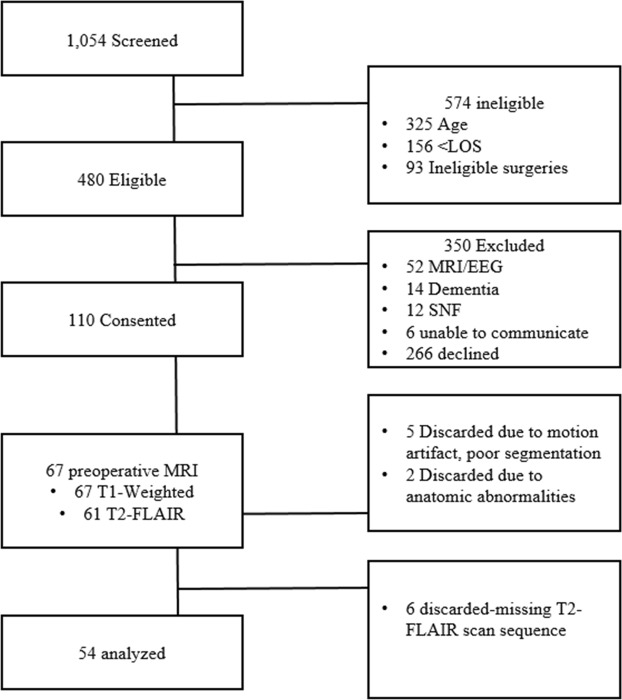
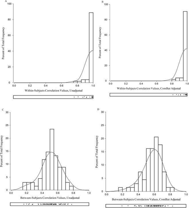
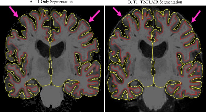
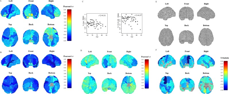
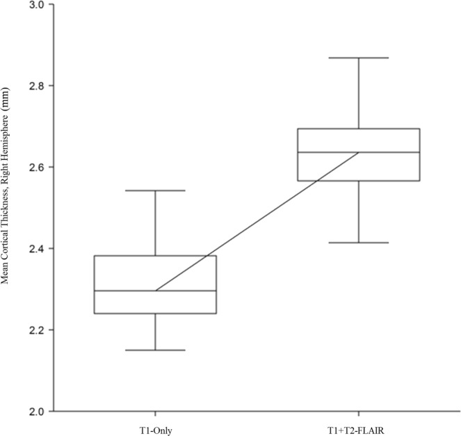
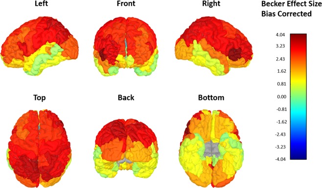
Cortical thickness is traditionally derived from T1-weighted MRI images. Recent studies have shown an improvement in segmentation with the combination of T1 + T2-FLAIR images. MRI data from 54 adults (mean: 71 years, 65-81 years, 48% females) that are part of an ongoing cohort study were analyzed to investigate whether T1 + T2-FLAIR cortical thickness measurements were superior to those derived from T1-weighted images in identifying age-related atrophy. T1-weighted and T2-FLAIR MRI images were processed through FreeSurfer v6.0. Data was extracted using the Desikan-Killiany (DKT) atlas. FreeSurfer's GUI QDEC examined age-related atrophy. Nonparametric tests, effect sizes, and Pearson correlations examined differences between T1-only and T1 + T2-FLAIR cortical thickness data. These analyses demonstrated that T1 + T2-FLAIR processed images significantly improved the segmentation of gray matter (chi-square x, p < 0.05) and demonstrated significantly thicker cortical thickness means (p < 0.05) with medium to large effect sizes. Significant regions of age-related cortical atrophy were identified within the T1 + T2-FLAIR data (FDR corrected, p < 0.05). This is in contrast to the T1-only data where no regions survived FDR correction. In summary, T1 + T2-FLAIR data were associated with significant improvement in cortical segmentation and the identification of age-related cortical atrophy. Future studies should consider employing this imaging strategy to obtain cortical thickness measurements sensitive to age-related changes.
皮质厚度传统上来源于 T1 加权 MRI 图像。最近的研究表明,T1+T2-FLAIR 图像的组合可提高分割效果。对来自一个正在进行的队列研究的 54 名成年人(平均年龄:71 岁,65-81 岁,女性占 48%)的 MRI 数据进行了分析,以研究 T1+T2-FLAIR 皮质厚度测量值是否优于 T1 加权图像,从而识别与年龄相关的萎缩。对 T1 加权和 T2-FLAIR MRI 图像进行了 FreeSurfer v6.0 处理。使用 Desikan-Killiany (DKT) 图谱提取数据。FreeSurfer 的 GUI QDEC 检查了与年龄相关的萎缩。非参数检验、效应量和 Pearson 相关性检验了 T1 加权和 T1+T2-FLAIR 皮质厚度数据之间的差异。这些分析表明,T1+T2-FLAIR 处理图像显著改善了灰质的分割(卡方 x,p<0.05),并显示出明显更厚的皮质厚度平均值(p<0.05),具有中等到大的效应量。在 T1+T2-FLAIR 数据中确定了与年龄相关的皮质萎缩的显著区域(经 FDR 校正,p<0.05)。这与 T1 加权数据形成对比,后者没有区域通过 FDR 校正。总之,T1+T2-FLAIR 数据与皮质分割的显著改善以及与年龄相关的皮质萎缩的识别相关。未来的研究应考虑采用这种成像策略来获得对年龄相关变化敏感的皮质厚度测量值。